Navigation auf uzh.ch
Navigation auf uzh.ch
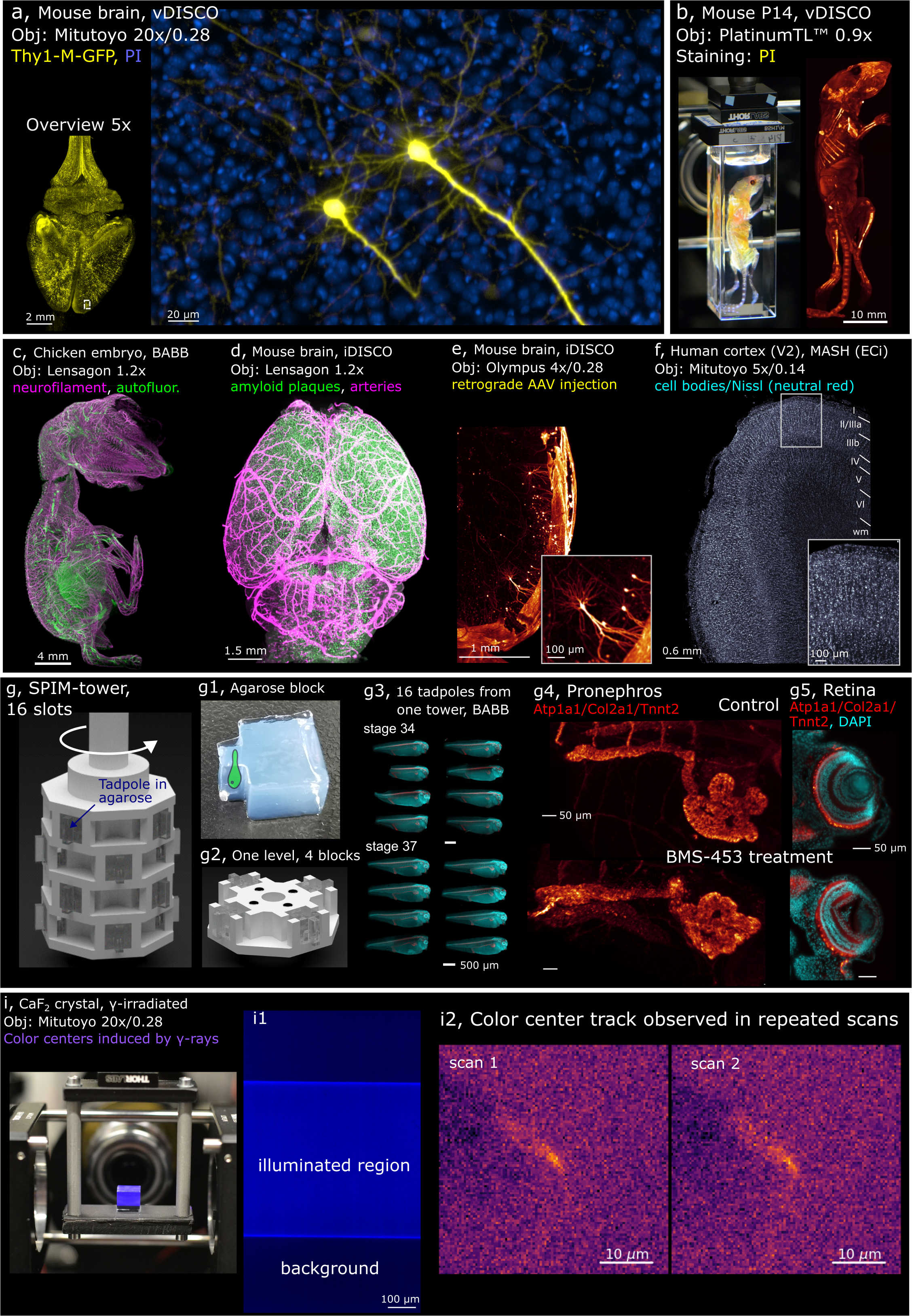
The mesoSPIM (mesoscale selective plane illumination microscopy) is the ideal instrument to quickly bridge scales from the µm- to the cm-level, which enables it to serve as an excellent tool for detailed three-dimensional anatomical investigations in many biological and medical fields (Voigt et al., 2019, Vladimirov et al, 2023).
Main features (upgrade 2023):
For further information: MesoSPIM.org
University Zurich, Irchel Campus, Room Y44-J-33.
Follow this link to apply for an introduction to the microscope.
The mesoSPIM excitation path is based on axially scanned light-sheet microscopy (ASLM). The waist of the light-sheet is translated through the sample in synchrony with the rolling shutter of the camera. The waist is translated with electrically tunable lenses by Optotune.
Light Sources and Lasers
The iChrome MLE comprises 3 diode lasers and 1 DPSS laser fully integrated in one compact housing.
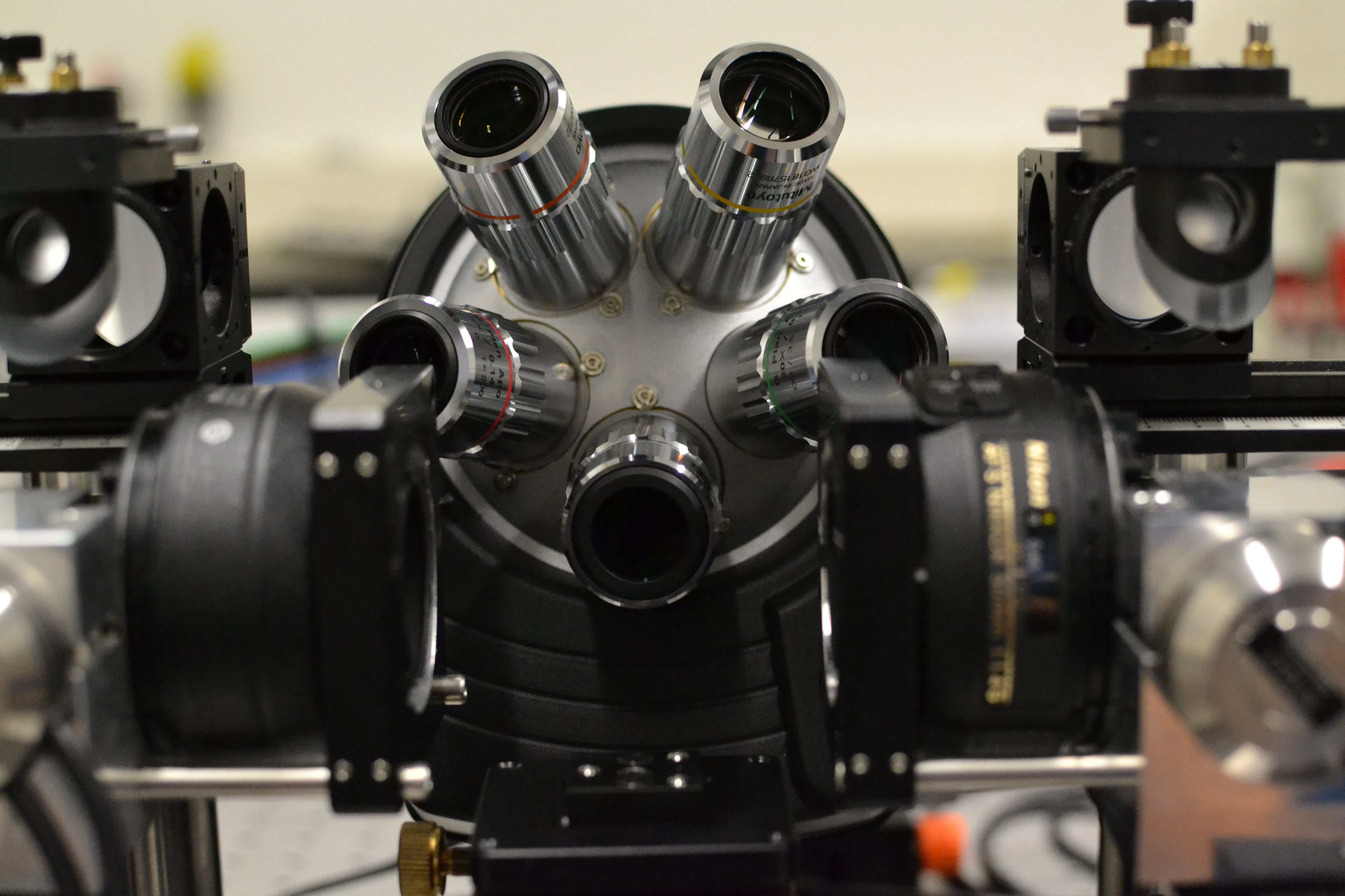
The mesoSPIM detection path consists of five long-working distance air plan apochromat objectives:
The microscope offers lateral (XY) resolution of 1.5-2.6 µm (depending on the objective) and axial (Z) resolution of 3.5 µm for all objectives, across a large field of view thanks to new-generation sCMOS camera Hamamatsu Orca Lightning.
Hamamatsu Orca Lightning sCMOS:
The effective pixel size in acquired image depends on objective magnification:
Pixel_size(image) = Pixel_size(sensor)/Magnification
| Magnification | Image pixel size, µm |
| 1x | 5.5 |
| 2x | 2.75 |
| 5x | 1.1 |
| 7.5x | 0.73 |
| 10x | 0.55 |
| 20x | 0.275 |
Field of view can be calculated from the sensor size and objective magnifications by the formula:
FOV(h,w) = Sensor(h,w)/Magnification
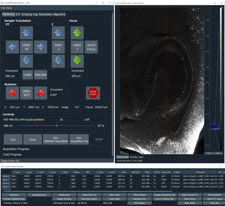
The microscope software, mesoSPIM-control, is developed by us. It is open-source and written in Python. It allows users to specify sequences of z-stacks using a table-based acquisition manager. The software can also be used to acquire large-scale tiling acquisitions.
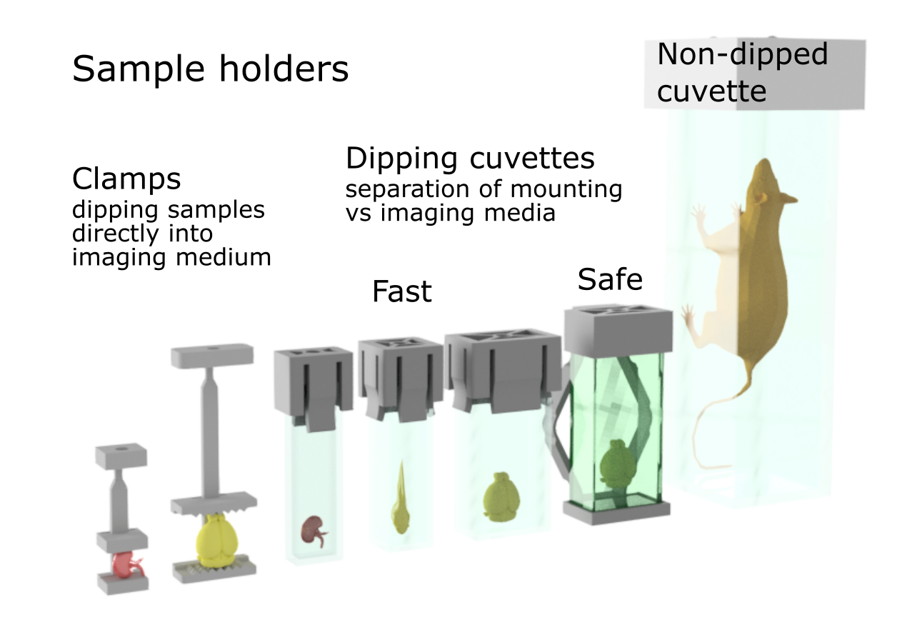
Depending on your sample, we have a variety of holders and imaging cuvettes.
Follow this link for further background information, documents and links.
Responsible Persons
If you have questions about the device please contact the responsible person.
Make sure to acknowledge the Center for Microscopy in your publication to support us.
How to acknowledge contributions of the Center for Microscopy