Navigation auf uzh.ch
Navigation auf uzh.ch
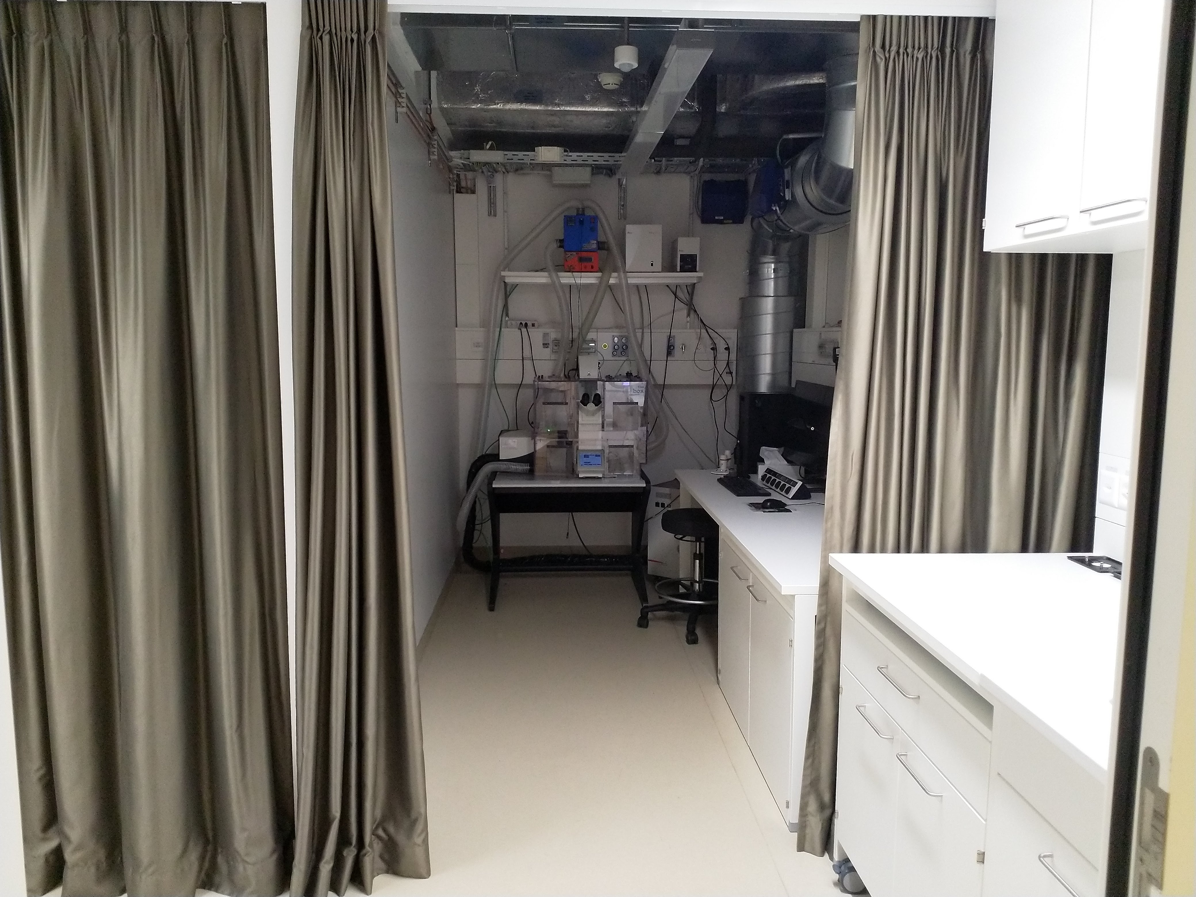
The Leica SP8 is an automated inverse confocal laser scanning microscope allowing simultaneous aquisition of 3 fluorescent channels and 1 transmitted light channel (BF or DIC).
University Zurich, Balgrist Campus, Lengghalde 5, 8008 Zurich, Room Microscopy W.112.
Follow this link to apply for an introduction to the microscope.
Dual scanning system:
Regular scanner: 10 - 1800 Hz
Resonant scanner: 8000 Hz
| Name | Magnification | NA | Immersion | WD (mm) |
|---|---|---|---|---|
| HC PL APO | 10x | 0.3 | Air | 11 |
| HC PL APO CS2 | 20x | 0.75 | Imm | 0.67 |
| HCX PL APO 37°C CS2 | 63x | 1.3 | Glycerol | 0.3 |
Two Hybrid (HyD) detectors, one photomultiplier tube (PMT) detector and one additional PMT for transmission. Excitation controlled by AOTF, beam splitters: RT 15/85, substrate, TD 488/552/638, DD 488/552 for separation between excitation and emission. Accurate emission detection by spectral detectors.
| Name | Excitation Range | Excitation Filter | Dichroic | Emission Filter |
|---|---|---|---|---|
| A4 | UV | BP 360/40 | 400 | BP 470/40 |
| GFP | blue | BP 470/40 | 500 | BP 525/50 |
| N2.1 | green | BP 515-560 | 580 | LP 590 |
| CFP | violet/blue | BP 436/20 | 455 | BP 480/40 |
| I3 | blue | BP 450/490 | 410 | LP 515 |
This microscope acquires images with improved axial resolution compared with more coventional widefield microscopes. In addition to the multicolor images the microscope can acquire large tile scans fully automatically. The microscope allows live imaging with controlled environmental conditions (CO2, temperature, humidity).
Follow this link for further background information, documents and links
Responsible Persons
If you have questions about the device please contact the responsible person.
Make sure to acknowledge the Center for Microscopy in your publication to support us.
How to acknowledge contributions of the Center for Microscopy