CLSM - Leica SP8 inverse (Irchel)
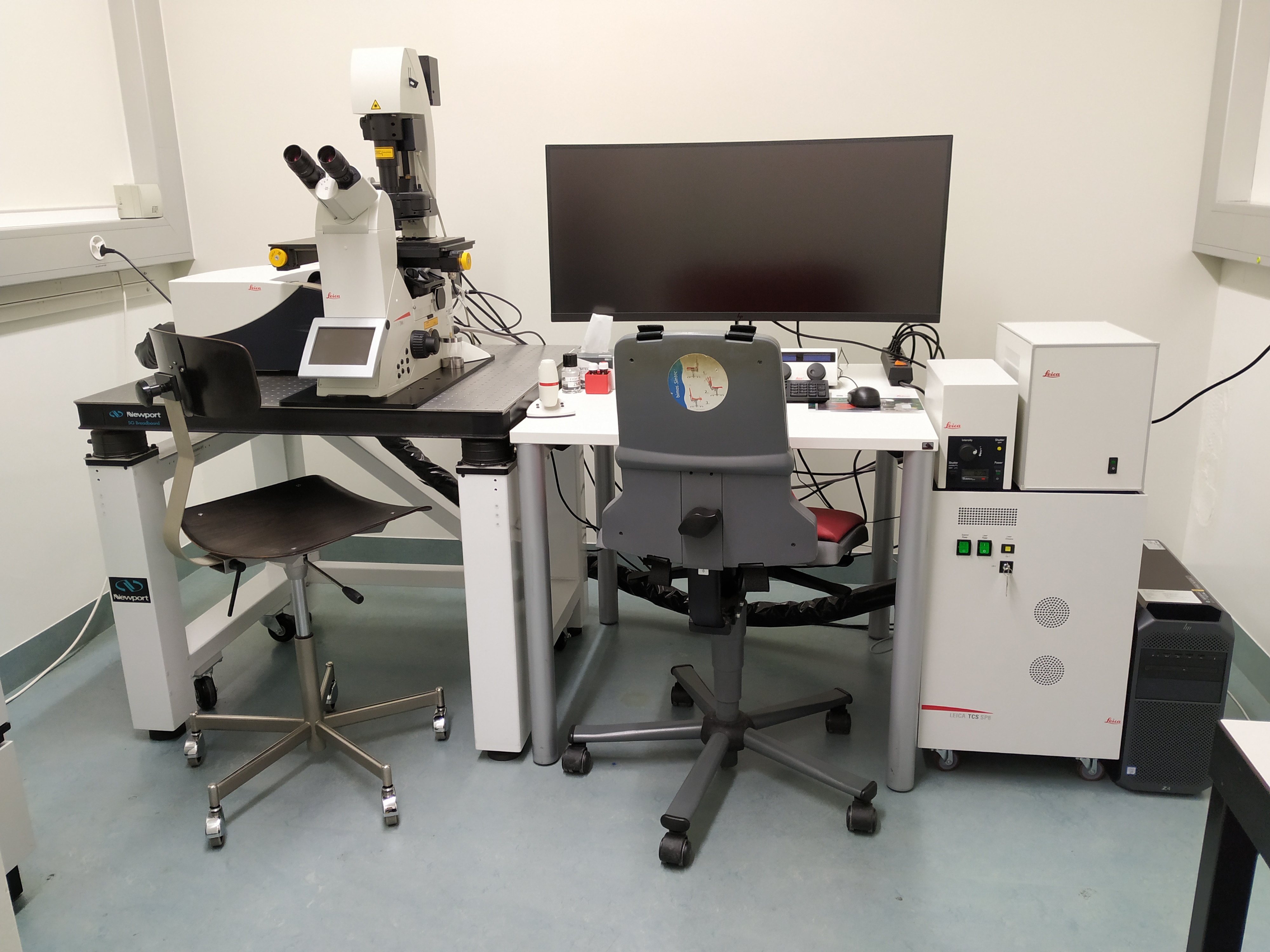
The Leica SP8 is an automated inverse confocal laser scanning microscope allowing simultaneous acquisition of 3 fluorescent channels and 1 transmitted light channel (BF). This system is ideal for fixed samples.
Location
University Zurich, Irchel Campus, Room Y55-K-41.
Training Request
Follow this link to apply for an introduction to the microscope.
Technical Specifications
Light sources and lasers
- Halogen lamp for transmitted light
- External fluorescence lamp LQ-HXP 120
- Solid state diode lasers: 405 nm (50 mW), 488 nm (20 mW), 552 nm (20 mW) and 638 nm (30 mW)
Scanners
Regular scanner: 10 - 1800 Hz
Objectives
| Name | Magnification | NA | Immersion | WD (mm) |
|---|---|---|---|---|
| HC PL APO | 10x | 0.3 | Air | 11 |
| HC PL APO CS2 | 20x | 0.75 | Imm | 0.67 |
| HC PL APO CS2 | 40x | 1.1 | Water | 0.65 |
| HCX PL APO CS2 | 63x | 1.4 | Oil | 0.14 |
CS2 objectives: improved color correction, perfect VIS-405; IMM = multi immersion (either water, glycerol or oil)
Detector System
Two Hybrid (HyD) detectors, one photomultiplier tube (PMT) detector and one additional PMT for transmission. Excitation controlled by AOTF, beam splitters: RT 15/85, substrate, TD 488/552/638, DD 488/552 for separation between excitation and emission. Accurate emission detection by spectral detectors.
Fluorescence Filters for widefield
| Name | Excitation Range | Excitation Filter | Dichroic | Emission Filter |
|---|---|---|---|---|
| DAPI | UV | BP 350/50 | 400 | BP 460/50 |
| GFP | blue | BP 470/40 | 495 | BP 525/50 |
| RHOD | green | BP 546/11 | 560 | BP 585/40 |
Make sure to acknowledge the Center for Microscopy in your publication to support us.
How to acknowledge contributions of the Center for Microscopy
Remarks
This microscope acquires images with improved axial resolution compared with more conventional widefield microscopes. In addition to the multicolor images in fixed samples the microscope can acquire large tile/overview scans and/or several positions fully automatically.
Literature and Links
Leica_Science_Lab
Further information (internal UZH use only)
Follow this link for further background information, documents and links
Responsible Persons
If you have questions about the device please contact the responsible person.
Make sure to acknowledge the Center for Microscopy in your publication to support us.
How to acknowledge contributions of the Center for Microscopy