CLSM - Leica Stellaris 5 inverse (USZ)
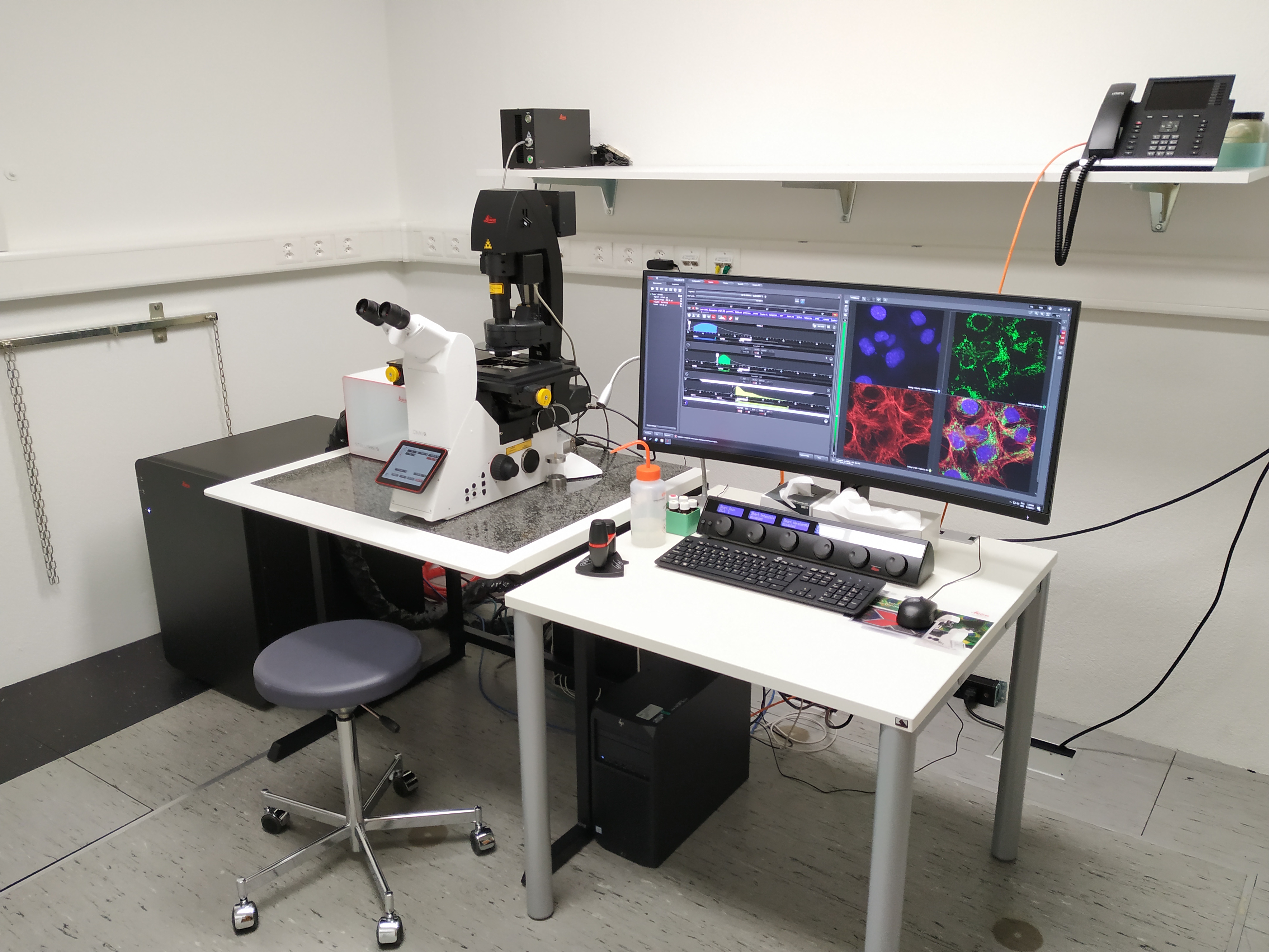
The Leica STELLARIS 5 is an automated inverse confocal laser scanning microscope allowing simultaneous acquisition of 3 fluorescent channels as well as a transmission bright field channel. The newest TauSense technology gives instant access to lifetime-based information, delivering additional insights to your experiments.This system is ideal for fixed samples.
Location
University Zurich Center, at the USZ Schmelzbergstrasse 12, Room PATH A25.
Training Request
Follow this link to apply for an introduction to the microscope.
Technical Specifications
Light sources and lasers
- Halogen lamp for transmitted light
- External fluorescence light source - Lumencor LED 3
- 405 nm diode laser
- Leica white light laser (WLL - 485 nm to 685 nm excitation)
Scanners
Regular scanner: 1 - 2600 Hz
Objectives
| Name | Magnification | NA | Immersion | WD (mm) |
|---|---|---|---|---|
| HC PL FLUOTAR | 5 | 0.15 | Air | 13.70 |
| HC PL APO CS2 | 10 | 0.4 | Air | 2.56 |
| HC PL APO CS2 | 20x | 0.75 | Air | 0.62 |
| HC PL APO CS2 | 20x | 0.70 | IMM | 0.26-0.17 |
| HC PL APO CS2 | 63x | 1.4 | Oil | 0.14 |
CS2 objectives: improved color correction, perfect VIS-405; IMM = multi immersion (either water, glycerol or oil)
Detector System
3 Hybrid (Power HyD S) detectors, and one additional detector for transmission (Trans PMT). Excitation controlled by AOTF. Accurate emission detection by spectral detectors.
Fluorescence Filters
| Name | Excitation Range | Excitation Filter | Dichroic | Emission Filter |
|---|---|---|---|---|
| DAPI. | UV | BP 405/60 | 455 | BP 470/40 |
| GFP | blue | BP 470/40 | 495 | BP 525/50 |
| RHOD | green | BP 546/10 | 560 | BP 585/40 |
Make sure to acknowledge the Center for Microscopy in your publication to support us.
How to acknowledge contributions of the Center for Microscopy
Remarks
This microscope acquires images with improved axial resolution compared with more coventional widefield microscopes.The newest TauSense technology gives instant access to lifetime-based information, delivering additional insights to your experiments. In addition to the multicolor images in fixed samples the microscope can acquire large tile scans fully automated thanks to the LAS X Navigator.
Literature and Links
Further information (internal UZH use only)
Follow this link for further background information, documents and links
Responsible Persons
If you have questions about the device please contact the responsible person.
Make sure to acknowledge the Center for Microscopy in your publication to support us.
How to acknowledge contributions of the Center for Microscopy