HCS - MD ImageXpress Confocal HT.ai
.jpg)
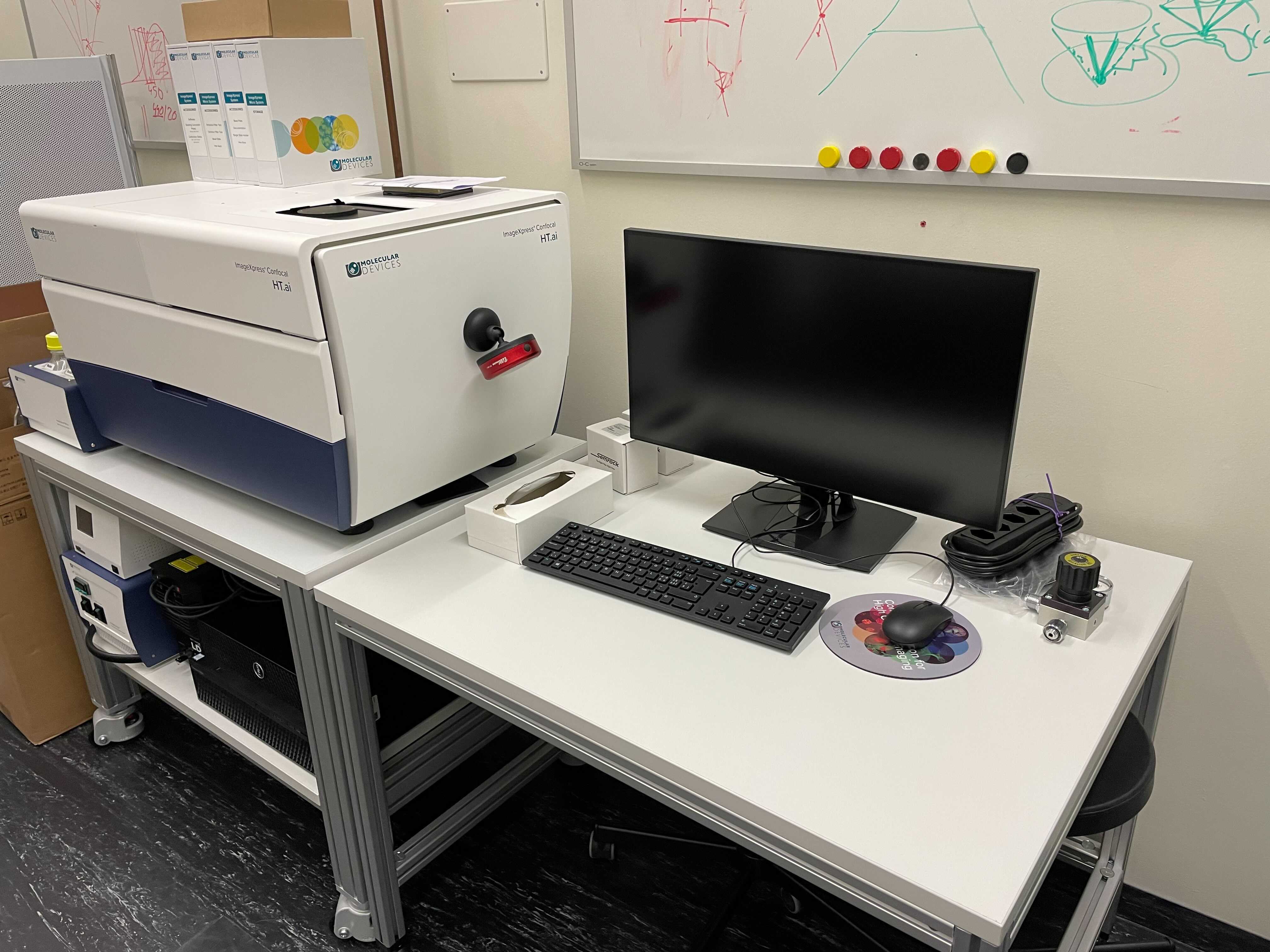
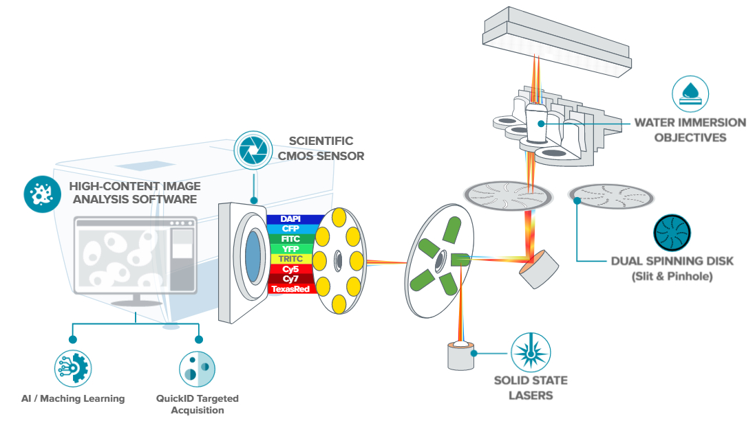
The ImageXpress Confocal HT.ai High-Content imaging system utilizes a seven-channel laser light source (410 nm - 750 nm) with eight imaging channels to enable highly multiplexed assays. Designed for experiments involving live cells as the spinning disk reduces phototoxicity and enables fast acquisition. The system supports slides and one to 1536-well microplates, round or flat bottom, low to high profile.
Please note: We provide assistance with optimization and troubleshooting exclusively for well plate–based carriers (polymer/glass bottom, 170 µm thickness, no foil-bottom plates). Unfortunately, we do not support slide or chambered slide formats, as these lack the robustness on this system required for reliable high-content imaging.
Location
University Zurich, Irchel Campus, Room Y42-H-91.
Training Request
Follow this link to apply for an introduction to the microscope.
Technical Specifications
Microscope
-
Inverted microscope (ImageXpress Confocal HT.ai)
-
Precision motorized X-Y (sample) stage
-
Dual disk unit allowing selection between 3 modes:
- 50 micron pinhole disk for your basic confocal requirements (better optical sectioning but less sensitive). Spacing between pinholes 500 um
-
50 micron (named 51 micron slit) for high throughput confocal requirements (faster acquisition but lower optical sectioning). Spacing between pinholes 250 um
- Widefield (not-confocal) imaging mode
Objectives
|
Default installed objectives |
||||||
|---|---|---|---|---|---|---|
| Name | Mag | NA | Immersion | WD (mm) | Pixel size (um/pixel) |
Recommended z step for deconvolution (um) |
| Nikon LWD Lambda S 40XC WI | 40x | 1.15 | Water | 0.61–0.59 |
0.1687 |
0.2 |
| Nikon CFI Apo LWD Lambda S 20XC WI | 20x | 0.95 | Water | 0.99–0.9 | 0.344 | 0.3 |
| Nikon CFI Plan Apo Lambda 10X | 10x | 0.45 | Air | 4 | 0.678 | |
| Nikon CFI Plan Apo Lambda 2X | 2x | 0.10 | Air | 8.5 | ||
| Optional objectives to be installed on request | ||||||
| Nikon CFI Plan Apo Lambda 20X | 20x | 0.75 | Air | 1 |
|
|
| Nikon CFI Plan Apo Lambda 40XC | 40x | 0.95 | Air | 0.25–0.17 |
Laser light source specifications
| Channel | Excitation Laser (nm) | Common Fluorophores | Emission Filter |
|---|---|---|---|
| DAPI | 405/20 | DAPI, Hoechst, Alexa Fluor 350, BFP | 452/45 nm |
| CFP | 445/20 | CFP | 483/32 nm |
| FITC | 470/21 | FITC, GFP, FAM, Alexa Fluor 488, BODIPY, Calcein, Fluo-4, MitoTracker Green | 520/28 nm |
| YFP | 520/10 | YFP | 562/40 nm |
| TRITC | 555 | Cy3, TRITC, Alexa Fluor 532 & 546 & 555, Rhodamine, TAMRA, LysoTracker Yellow, MitoTracker Orange |
598/25 nm |
| Texas Red | 555 | Texas Red, mCherry, Alexa Fluor 568 & 594, Calcium Crimson, Cy3.5, HcRed, MitoTracker Red, Propidium Iodide | 624/40 nm |
| Cy5 | 638/17 | Cy5, Alexa Fluor 647 & 660, DRAQ5, APC (Allophycocyanin), BODIPY 650/665, DiD, SYTO Red, TOTO-3, and TO-PRO-3 | 692/40 nm |
| Cy7 | 725/30 | Cy7 | 794/32 |
Camera Systems
- High-quantum efficiency 16-bit, >4 megapixel scientific CMOS sensor
Accessories
- Incubation system for live cell imaging (ImageXpress)
- Allows for a variety of experimental protocols with 0 - 20 % CO2, 0 - 100% nitrogen, 0 - 100% Oxygen.
- IN Carta Image Analysis Software
Responsible Persons
If you have questions about the device please contact the responsible person.
Make sure to acknowledge the Center for Microscopy in your publication to support us.
How to acknowledge contributions of the Center for Microscopy
Recommended imaging supports
In order to ensure reliable imaging at this instrument we recommend using imaging supports with high performance #1.5 cover glass or equivalent. The following imaging supports have been successfully used:
Plate |
Ref | plate definitions file (*.plt) |
|---|---|---|
| Cellvis P12-1-5H glass bottom | P12-1.5H-N | P12-1.5H-N (PLT, 1 KB) |
| Cellvis P96-1-5H-N | P96-1.5H-N | P96-1.5H-N (PLT, 2 KB) |
| ibidi 96 well plate ibidi glass | #1.5 glass bottom, Cat.No:89627 | 89627 |
| ibidi 96 well plate | #1.5 glass-like polymer coverslip bottom, Cat.No:89626 | 89626 (PLT, 2 KB) |
| ibidi 384 well plate glass | #1.5 glass bottom, Cat.No:88407 | 88407 (PLT, 1 KB) |
If you'd like to ask for a free sample to test, please use the link https://ibidi.com/content/875-ibidi-free-sample-program
We do not support or troubleshoot autofocus strategies using ultra-thin transparent film bottoms.
Please contact us in case you have further question.