Spinning Disk - Visitron CSU-W1 (Irchel)
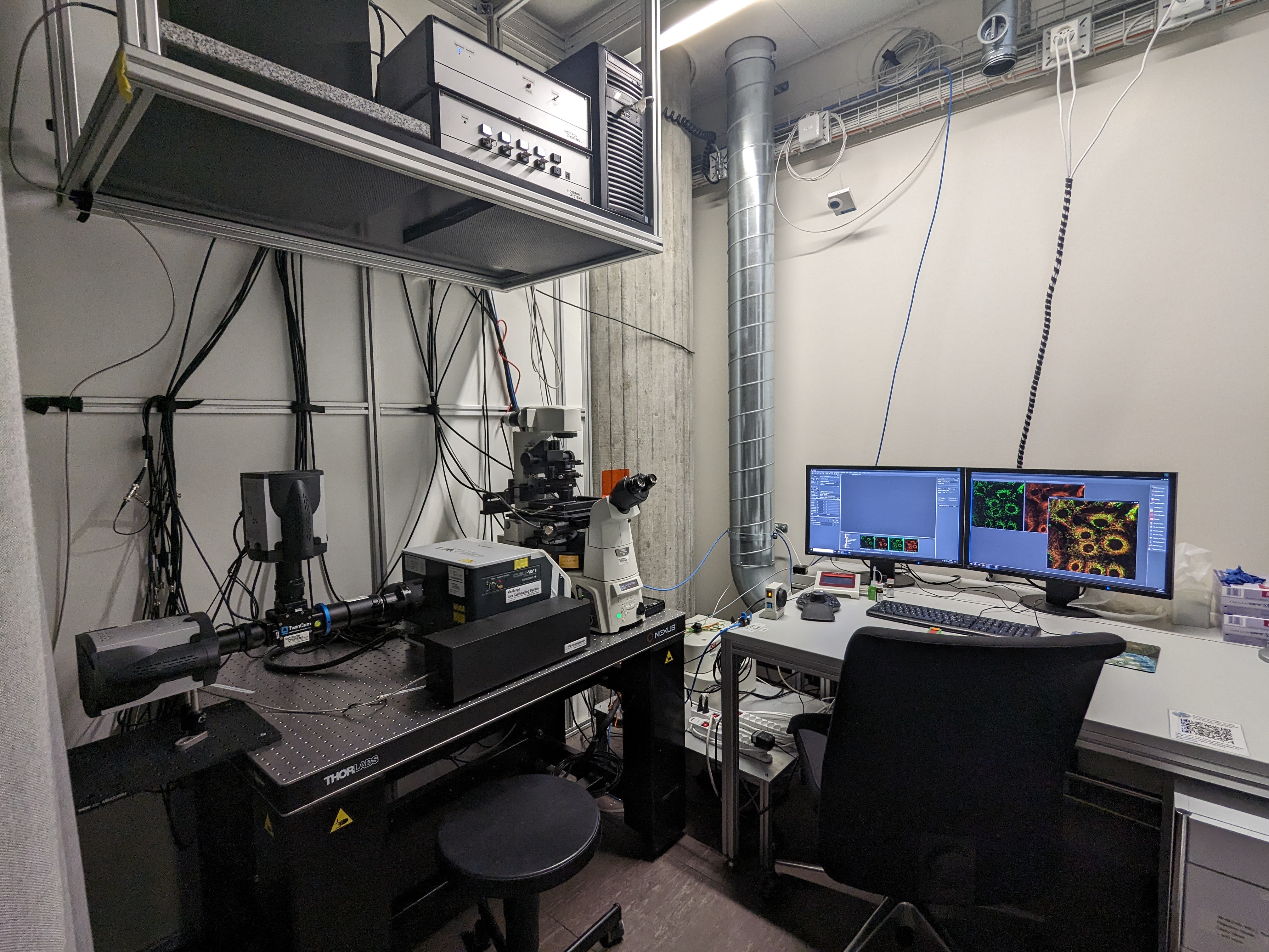
The Spinning Disk Microscope is a high-end real time confocal imaging system, equipped with the Yokogawa Confocal scanning unit W1 (2 sets of spinning disks for low and high resolution imaging).
It is further equipped with two EMCCD cameras, which allow for fast and sensitive simultaneous acquisition of two-color (live) samples, also well suited for low fluorescence emission.
The spinning disk facilitates long time investigations (hours to days) of cells and tissue, showing reduced phototoxicity and bleaching, while the Perfect Focus System (PFS, active focus control) maintains the focus.
A stage top incubation system allows control of temperature and C02 levels enabling live cell experiments.
Location
University Zurich, Irchel Campus, Room Y24-F-14.
Training Request
Follow this link to apply for an introduction to the microscope.
Technical Specifications
Microscope
- inverted microscope (Nikon Eclipse Ti-E)
- Yokogawa Spinning Disc System W1 (2 discs: 25um pinholes for low NA objectives and 50um pinholes for high NA objectives)
- motorized xyz stage
- perfect focus system (PFS)
- highly precise motorized z-focus/nosepiece
Light Sources and Lasers
- high-power LED illumination system: selection of spectral lines for DAPI, GFP, mCherry and Cy5
- cooled LED (525nm) for transmission light
- solid state diode lasers: 405 nm (120 mW), 488 nm (100 mW), 561 nm (200 mW), 640 nm (150 mW)
Objectives
| Name | Magnification | NA | Immersion | WD (mm) |
|---|---|---|---|---|
| CFI PlanApo | 10x (DIC) | 0.45 | Air | 4.0 |
| CFI S PlanFluor ELWD | 40x (Ph) | 0.6 | Air | 2.8-3.6 |
| CFI S PlanFluor ELWD | 20x (Ph) | 0.45 | Air | 6.9-8.2 |
| CFI PlanApo VC | 60x (DIC) | 1.4 | Oil | 0.13 |
| CFI PlanApo | 100x (DIC) | 1.4 | Oil | 0.13 |
| CFI SR HP PlanApo Lambda S | 100x (DIC) | 1.35 | Silicon oil | 0.28-0.31 |
| Additional Objective - not installed | ||||
| CFI PlanFluor | 20x (DIC) | 0.75 | IMM | 0.33-0.35 |
IMM = multi immersion (either water, glycerol or oil)
Fluorescence Filters
Beam path

Filter for confocal imaging (implemented in CSU-W1)
Mainly used if 1 camera imaging is desired. All images acquired with CAM1.
| Laser Line | Excitation Range | Dichroic (DM 1) | Emission filter (FW) | TwinCam Unit |
|---|---|---|---|---|
| 405 | UV | 410 | BP 460/50 | empty (no DM2, no EM1) |
| 488 | blue | 504 | BP 525/50 | empty (no DM2, no EM1) |
| 561 | green | 582 | BP 609/54 | empty (no DM2, no EM1) |
| 640 | red | 669 | BP 700/75 | empty (no DM2, no EM1) |
Filter for confocal imaging (implemented in TwinCam)
Mainly used if 2 camera imaging is desired.
| Laser Lines | FW | Image Splitter (DM 2) | EM /Camera 1 | EM /Camera 2 |
|---|---|---|---|---|
| 405/561 | open | A561 LP |
BP 605/52 (or BP 595/33) |
BP 460/50 |
| 405/640 | open | A561 LP | BP 700/75 | BP 460/50 |
| 488/561 | open | A561 LP | BP 605/52 (or BP595/33) | BP 525/50 |
| 488/640 | open |
A561 LP or A647 LP |
BP 700/75
|
BP 525/50 |
| 561/640 | open | A647 LP | BP 700/75 |
BP 595/33 BP 605/52 (only for TwinCam single) |
Filter for Epi-Fluorescence (implemented in LED housing and microscope stativ)
| LED (Excitation range) | Excitation Filter | Dichroic | Emission Filter | FW |
|---|---|---|---|---|
| DAPI (UV) | BP 390/18 | acc. QUAD | acc. QUAD | BP 460/50 |
| GFP (blue) | BP 485/20 | acc. QUAD | acc. QUAD |
BP 525/50 |
| mCherry (green) | BP 560/25 | acc. QUAD | acc. QUAD | BP 609/54 |
| Cy5 (red) | BP 650/13 | acc. QUAD | acc. QUAD | BP 700/75 |
| QUAD (UV/blue/green/red) |
BP 387/18; 485/30; 559/30; 649/22 |
410; 504; 582; 669 |
BP 440/50; 521/30; 607/40; 700/60 |
The QUAD Filter cube is only used for Epi-Fluorescence imaging. In confocal mode, filters in the microscope stand are automatically removed from the light path. For dual Cam imaging, FW needs to be open, and the appropriate EM 1/2 as well as corresponding DM 2 have to be inserted into the TwinCam.
For further imaging methods/illumination pathway options please contact the responsible person.
Camera Systems
- 2x iXon-888Life Back-illuminated EMCCDs
- 1024 x 1024 pixel (13 x 13 um pixel size)
- fastest read out speed in full FOV: 26fps
Accessories
- Incubation system for live cell imaging (Oko Lab technology) - stage top incubator
-
Several heating insert bottom plates:
- 2 x 35mm petri dishes, 1 x 1"x2" chambered cover glass
- multiwell plates
- Objective heaters for different lense diameters
Literature and Links
Further information (internal UZH use only)
Follow this link for further background information, documents and links.
Responsible Persons
If you have questions about the device please contact the responsible person.
Make sure to acknowledge the Center for Microscopy in your publication to support us.
How to acknowledge contributions of the Center for Microscopy