Multiphoton - Custom Built Microscope (Irchel)
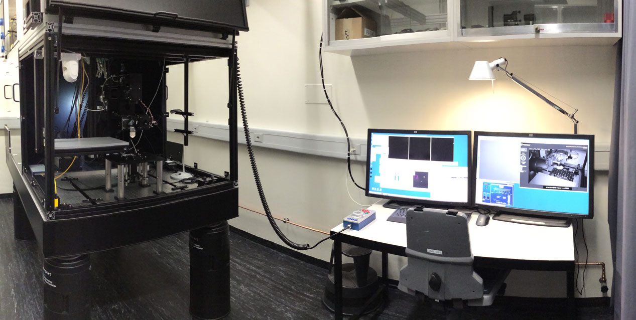
In corporation with Prof. Bruno Weber we custom-built a highly versatile multiphoton microscope. The microscope was financed by the UZH and NCCR Kidney to foster the in vivo imaging of kidney function and diseases (NCCR Kidney: Prof. Andrew Hall) as well as other research topics. The microscope allows full 360 degrees rotational freedom of the objective and large x, y, z movements. It is equipped with an advanced laser system with a very broad tuning range allowing deep tissue imaging and the excitation of red fluorescent dyes, which previously were not usable in multiphoton imaging.
Location
University Zurich, Irchel Campus, Room Y42-H-89
Training Request
The microscope is open for everybody, but dependent on usage, some priorities are given to the research group of Prof. Andrew Hall. If you have questions regarding the microscope or want to discuss your research project, please directly contact Joana Delgado Martins.
Technical Specifications
Light Sources and Lasers
- Chameleon Discovery NX with TPC: Dual (upgraded 2025)
Chameleon Discovery delivers high peak power right to the sample plane using built in dispersion precompensation for the tunable output. The combination of short pulses (100fs at 900nm), high average power (>2.7 W at 900nm) and wide tuning range (660-1320nm) gives brighter and deeper images for in-vivo imaging applications.
Repetition Rate (MHz) 80 ±0.5
A second output at 1040nm, with high power (>2800mW) and short pulses (<140fs) further enables multimodal capability for dual color excitation and activation of optogenetics probes.
Total Power Control (TPC) provides fast rise time (< 500 ns) and high extinction ratio (>1000:1) modulation of both output with guaranteed pristine beam quality which can be delivered directly a two photon microscope.
Objectives
| Name | Magnification | NA | Immersion | WD (mm) |
|---|---|---|---|---|
| Olympus XLPlan N ( XLPLN25XWMP2) | 25x | 1.05 | Water | 2.0 |
| Zeiss W Plan-Apochromat | 20x | 1.0 | Water | 2.3 |
Objective Adapters
To mount microscope objectives from different manufacturers, the following objective adapters are available:
| Thread | Description |
|---|---|
| M25x0.75 | For Olympus XLPlan N and other objectives |
| M27x0.75 | For Zeiss W Plan-Apochromat and other objectives |
| M32x0.75 | Special objectives |
| 1.035”-40 | Thorlabs SM1 |
| 26x0.706 | For Olympus objectives |
| 0.8”x1/36” | RMS (Zeiss and Olympus objectives) |
Fluorescence Filters
| Name | Dichroic | IR Block | Detector 1 | Detector 2 |
|---|---|---|---|---|
| Cube 1 | 506 nm | 680 nm SP | 475/50 BP | 542/50 BP |
| Cube 2 | 560 nm | 770 nm SP | 520/70 BP | 607/70 BP |
| Cube 3 | 580 nm | 770 nm SP | 535/50 BP | 630/69 BP |
Camera System
IDS 1540LE CMOS monochrome camera, 25 fps, 1280x1024 pixels. The camera is used for aligning the instrument and for finding the focus. Fluorescent imaging is not possible.
Accessories
- Custom made, heated mouse holder for inverted in-vivo experiments
- Anesthesia system for small rodents
- Binocular for surgery of small animals
- Two custom made microscope stages. An upright stage with transmission LED for imaging microscope slides and tissue slices in perfusion chambers. An inverted Stage for in-vivo experiments
- Micromanipulator for physiology experiments
Remarks
- Laser safety is a very important aspect in operating custom built two-photon microscopes. The infrared laser used in this system is a class 4 laser. To reduce the risk of injury various technical and organizational measures have been taken. Therefore, access to the microscope is highly restricted and only well trained and experienced users are allowed to operate the system. It is compulsory to understand and accept laser safety measures and rules by signing the laser safety regulations before access to the system is given.
- The microscope containment is completely built using industrial aluminum profiles. This allows easy adaptions and expansion according to experimental needs.
Literature and Links
Further information (internal UZH use only)
Follow this link for further background information, documents and links.
Responsible Persons
If you have questions about the device please contact the responsible person.
Make sure to acknowledge the Center for Microscopy in your publication to support us.
How to acknowledge contributions of the Center for Microscopy