Widefield - ZEISS Elyra 7 Lattice SIM² (Irchel)

ZEISS Elyra 7 Lattice SIM²
The Zeiss Elyra 7 with different super-resolution modalities, allows detailed visualization of samples beyond the diffraction limit of conventional light microscopy. With its 2 fast sCMOS cameras, this system is able to generate multiple color data simultaneously at incredibly high frame rates.
Responsible Person |
|
Location |
University Zurich, Irchel Campus, Room Y42-H-79. |
Training Request |
Follow this link to apply for an introduction to the microscope |
The Elyra 7 features the following modalities:
Widefield, DIC |
Widefield (WF) mode (sample illumination with arc lamp), Laser WF mode (sample illumination with laser) |
Lattice SIM |
Structured Illumination Microscopy, allowing fast and gentle super-resolution imaging (~120 nm in xy and ~300 nm in z) in 3 dimensions. Lateral resolution (XY): 120 nm, axial resolution (Z): 300 nm (typical experimental FWHM values with objective lens Plan-Apochromat 63× / 1.40 Oil DIC, subresolution beads of 40 nm diameter and excitation at 488 nm) |
Lattice SIM2 |
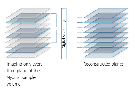
SIM² is a novel, image reconstruction algorithm by ZEISS that increases the resolution and sectioning quality of structured illumination microscopy data. Unlike conventional reconstruction algorithms, SIM² is a two-step image reconstruction algorithm. First, order combination, denoising and frequency suppression filtering are performed. All the effects resulting from these digital image manipulations are translated into a digital SIM point spread function (PSF). The subsequent iterative deconvolution uses this very PSF. Similar to advantages of using experimental PSF for deconvolution of hardware-based microscopy data, the SIM² algorithm is superior to conventional one-step image reconstruction methods in terms of resolution, sectioning and robustness. The new SIM2 module doubles the conventional SIM resolution and achieves up to ~60 nmlaterally and ~200 nm axially. The Lattice SIM illumination allows for a better signal to noise ratio, more gentle imaging and can reach up to 255 fps during time lapse acquisition, well suited to capture highly dynamic biological processes in live samples. It further allows deeper imaging of samples up to around 70 µm thickness in comparison to only 20 µm of sample thickness in classical SIM. The speed of this imaging modality is well suited to capturing dynamic biological processes in live samples (significantly faster than regular laser scanning confocal). Lateral resolution (XY): down to 60 nm, axial resolution (Z): down to 200 nm (typical experimental FWHM values with objective lens Plan-Apochromat 63× / 1.40 Oil DIC, subresolution beads of 40 nm diameter and excitation at 488 nm; Resolution is sample and SNR dependent.) |
Apotome |
Grid-based optical sectioning to create highly contrasted images with high lateral and axial resolution. The Apotome mode is also amenable to SIM2 processing, allowing for high speed acquisition with high contrast, high resolution and low phototoxicity. In combination with SIM2 module, apotome imaging can get lateral rsoltuion down to ~110 nm and axial resolution to ~300 nm. ApoTome imaging is also faster than classic SIM and laser scanning microscopes, allowing for time lapse imaging of faster cellular/tissue dynamics. Lateral resolution (XY) of 140 nm, axial resolution (Z) of 275 nm for 40×; lateral resolution (XY) of 285 nm, axial resolution (Z) of 550 nm for 20×; lateral resolution (XY) of 710 nm, axial resolution (Z) of 1300 nm for 10× |
SMLM |
Single-molecule localization microscopy like dSTORM, PALM and PAINT allowing resolution down to ~20-30 nm in xy and ~50-80 nm in z. |
HILO & TIRF |
Highly inclined and laminated optical sheet (HILO) and total interbal reflection illumination (TIRF). |
Technical Specifications
Microscope body |
|
||||||||||||||||||||||||||||||||||||||||||
Light Sources and Lasers |
|
||||||||||||||||||||||||||||||||||||||||||
Camera System |
|
||||||||||||||||||||||||||||||||||||||||||
Environmental control |
|
||||||||||||||||||||||||||||||||||||||||||
Accessories |
|
||||||||||||||||||||||||||||||||||||||||||
Available Optics |
|
||||||||||||||||||||||||||||||||||||||||||
Available Filters |
|
||||||||||||||||||||||||||||||||||||||||||
Sample requirements |
|
||||||||||||||||||||||||||||||||||||||||||
Acquisition speed (Lattice SIM) |
|
||||||||||||||||||||||||||||||||||||||||||
Acquisition speed (SIM Apotome mode) |
|
||||||||||||||||||||||||||||||||||||||||||
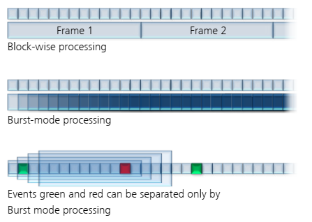
Leap mode and Burst mode |
|
||||||||||||||||||||||||||||||||||||||||||
User Guide |
|||||||||||||||||||||||||||||||||||||||||||
Links & Literature |
ZEISS Dual Iterative SIM Sample preparation for ELYRA 7 (SIM & SMLM) ibiology - Super-Resolution: Localization Microscopy (Bo Huang) |
.2022-12-09-12-13-40.png)
Make sure to acknowledge the Center for Microscopy and Image Analysis in your publication to support us.
How to acknowledge contributions of the Center for Microscopy and Image Analysis