Widefield - GE IN Cell Analyzer 2500HS (Irchel)
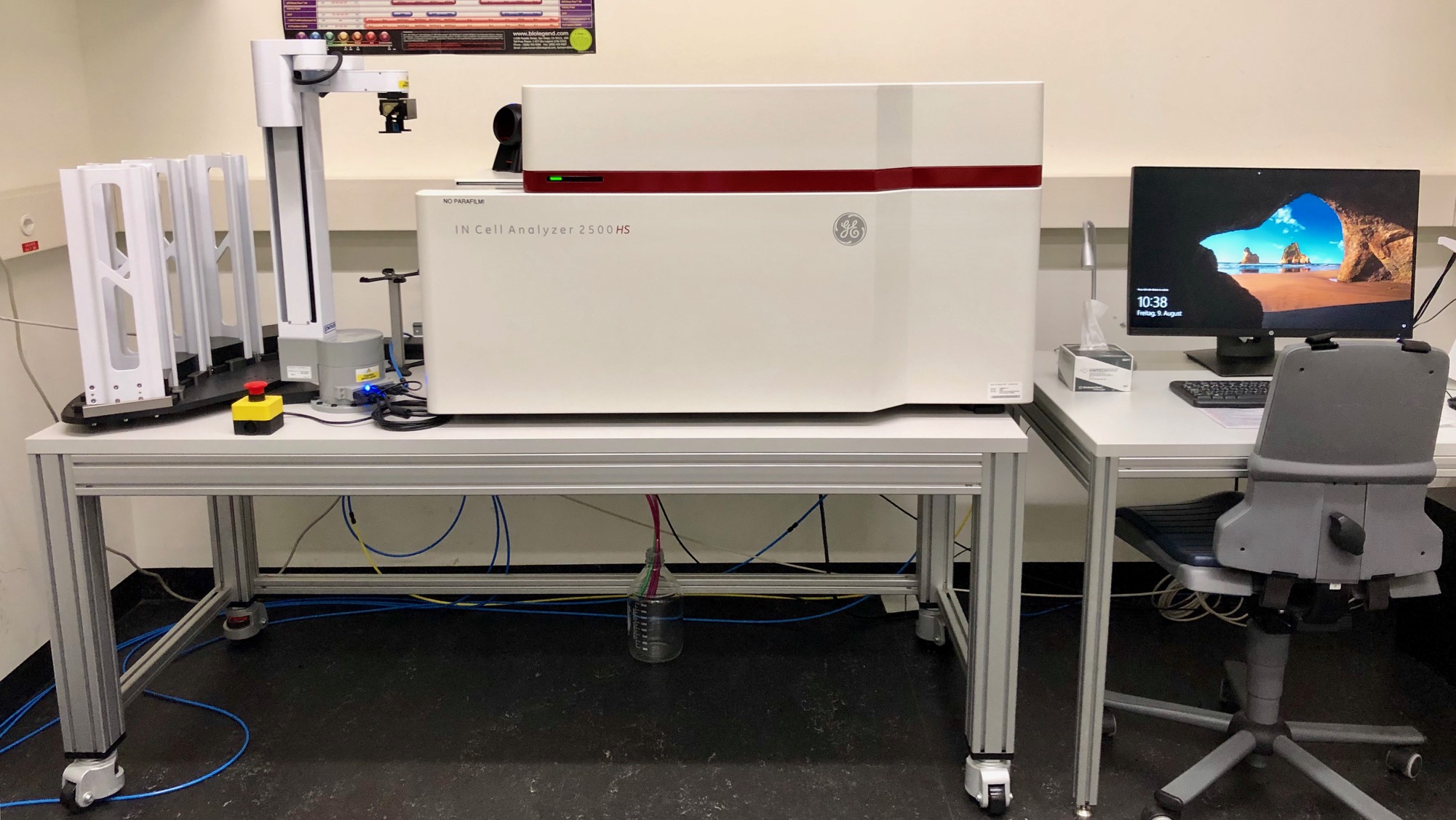
The INCell Analyzer 2500HS is a fully-automated widefield fluorescence imaging system dedicated to life cell imaging and high content and high throughput imaging. The system allows for sample automation using a robot as well as for liquid handling for drug applications and water immersion.
Location
University Zurich, Irchel Campus, Room Y42-H-91.
Training Request
Follow this link to apply for an introduction to the microscope.
Technical Specifications
Microscope
- GE inverted microscope.
Light Sources
- Seven-color Solide State Illuminator (SSI) for fluorescence excitation
- White LED for transmitted light
| Name | Magnification | NA | Immersion | WD (mm) |
|---|---|---|---|---|
| CFI Plan Apo Lambda | 10x | 0.45 | Air | 4.0 |
| CFI Plan Apo Lambda | 20x | 0.75 | Air | 1.0 |
| CFI Plan Apo Lambda | 40x | 0.95 | Air | 0.2-0.11 |
|
CFI Plan Apo Lambda* |
40x | 1.15 | Water | 0.61 |
*Automated water immersion system is now fully supported in InCell software.
Fluorescence Filters
IN Cell Analyzer 2500HS is equipped with two quad band pass polychroic mirrors and single band pass emission filters.
| Polychroic beam splitter BGOFR_1 | ||
|---|---|---|
|
Filter name |
Excitation | Emission |
| Blue | BP 390/18 | BP 432.5/47 |
| Green_1 | BP 475/28 | BP 511.5/23 |
| Orange | BP 542/27 | BP 587.5/47 |
| FarRed | BP 632/22 | BP 676/48 |
| Polychroic beam splitter BGRFR_2 | ||
|---|---|---|
|
Filter name |
Excitation | Emission |
| Blue | BP 390/18 | BP 432.5/47 |
| Green_2 | BP 475/28 | BP 526/52 |
| Red | BP 575/25 | BP 607.5/19 |
| FarRed | BP 632/22 | BP 676/48 |
Camera Systems
- PCO - sCMOS, 16 bit, 2048 x 2048 pixel, pixel size 6.5 µm, readout speed: 272 MHz
Imaging Modes
- Transmitted light, DIC (digital), Phase (digital)
- 2D deconvolution, Advanced 2D deconvolution, 2.5D, 3D deconvolution
Accessories
-
Complete environmental control (temperature, CO2, N2, humidity)
- Sample holders for multiwell plates, regular slides (1 or 4 pieces), chambered coverglass slides, 35 mm dishes
- liquid handling
- robotic automation
Remarks
Templates for slides
In order to facilitate the imaging process of slides with the GE IN CellAnalyzer we recommend to prepare the slides according to the templates depicted in the following document GE templates (PDF, 308 KB). For each of these slide-setups you will find a predefined slide profile in the GE software.
Literature and Links
Follow this link for further background information, documents and links (internal UZH use only).
Responsible Persons
If you have questions about the device please contact the responsible person.
Make sure to acknowledge the Center for Microscopy in your publication to support us.
How to acknowledge contributions of the Center for Microscopy