Widefield - Nikon Ti2 (Balgrist Campus)
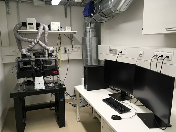
This inverted widefield system is suited to image live cells as well as fixed fluorescence and histologically stained samples. A temperature and CO2 controlled environment is possible for live cell experiments.
Location
University Zurich, Balgrist Campus, Lengghalde 5, 8008 Zurich, Room Microscopy W.112
Training Request
Follow this link to apply for an introduction to the microscope.
Technical Specifications
Microscope
Nikon Eclipse Ti2, inverted
Light Sources
- Lumencor sola light engine
| Name | Magnification | NA | Immersion | WD (mm) |
|---|---|---|---|---|
| Plan Fluor PhL DL | 4x | 0.13 | Air | 16.5 |
| Plan Fluor Ph1 DL | 10x | 0.3 | Air | 5.2 |
| Plan Apo l Ph2 DM | 20x | 0.75 | Air | 1.0 |
| S Plan Fluor ELWD DIC N1 | 40x | 0.6 | Air | 3.6 |
| Plan Fluor DIC H N2 | 40x | 1.3 | Oil | 0.24 |
| Plan Apo VC WI DIC N2 | 60x | 1.2 | Water | 0.31 |
Ph = phase contrast, DL = Dark Low Contrast, DM = Dark Medium Contrast, DIC = Differential Interference Contrast
Fluorescence Filters
| Name | Excitation | Dichroic | Emission |
|---|---|---|---|
| DAPI | BP 377/50 | 409 | BP 447/60 |
| GFP | BP 466/40 | 495 | BP 525/50 |
| YFP | BP 500/24 | 520 | BP 542/27 |
| Cy3 | BP 531/40 | 562 | BP 593/40 |
| Cy5 | BP 628/40 | 660 | BP 692/10 |
Camera Systems
-
cooled Nikon monochrome fluorescent DS-Qi2 CMOS camera
4908 x 3264 pixel (16.25 MP)
pixel size 7.3 um
25 mm FOV
Frame rate: 6 fps (4908×3264) up to 45 fps (1636×1088)
- Nikon Colour DS-Ri2, CMOS
Pixel: 4908 x 3264 pixel (16.25MP)
Pixel size: 7.3 µm
16bit
Frame rate: 6 fps (4908×3264) up to 45 fps (1636×1088)
Accessories
- IR Hardware Autofocus (Nikon Perfect Focus System - PFS)
- Temperature and CO2/O2 control (Okolab bold line)
- NIS-Elements with JOBS module (enabling e.g. automatic screening of 96 or 384 well plates)
Responsible Persons
If you have questions about the device please contact the responsible person.
Make sure to acknowledge the Center for Microscopy and Image Analysis in your publication to support us.
How to acknowledge contributions of the Center for Microscopy and Image Analysis