Widefield - Leica Thunder Imager 3D Live Cell (Irchel)
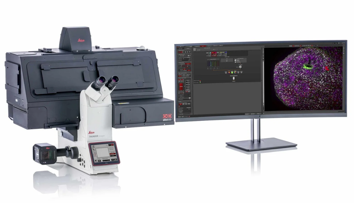
The LeicaTHUNDER Imager 3D Live Cell on the Leica DMi8 platform – a fully motorized, inverted research microscope equipped with hardware-based adaptive focus control (AFC) for precise drift correction and rapid focusing in multi-well applications. This versatile instrument is designed to capture fixed and live samples in up to 8 channels with remarkable speed. With THUNDER - Computational Clearing methodology, images are instantly deconvolved, offering confocal-like clarity while excelling in rapid time-lapse recording and minimizing phototoxicity. Built on the reliable DMi8 platform, it accommodates various sample types with stage holders for slides, petri dishes, and tissue culture plates. Imaging is acquired via a suite of high-NA objectives ranging from 5X to 100X. Two available cameras provide multi-channel fluorescence imaging via a high-sensitivity sCMOS B/W camera (Leica monochrome fluoroscence DFC9000 GTC) or high-resolution color imaging via a 12 bit CMOS RGB camera (Leica digital color camera DMC6200). Rapid switching between fluorescent dye channels is augmented by the use of a single quad-cube excitation filter set coupled with an external emission filter wheel. Experience enhanced live cell imaging capabilities, including precise focus maintenance over extended acquisition periods.
Responsible Person |
|
Location |
University Zurich, Irchel Campus, Room Y42-H-93. |
Training Request |
Follow this link to apply for an introduction to the microscope |
The Thunder Imager features the following modalities:
Widefield |
Widefield (WF) mode (sample illumination with LED) |
DIC |
Differential Interference Contrast (DIC) microscopy is another technique used to enhance contrast in transparent samples. It works by splitting a beam of light into two, altering their paths slightly, and then recombining them. This creates an interference pattern, making subtle variations in the sample's refractive index more visible. DIC microscopy produces images with three-dimensional appearance and enhanced contrast, making it useful for studying living cells and tissues in great detail. |
Phase contrast |
Phase contrast is a microscopy technique that makes transparent samples visible by enhancing the differences in how light waves pass through them. It helps to reveal details in objects like cells or tiny organisms that are hard to see with regular microscopy. |
Fura-2 |
Fura-2 imaging is a method in cell biology for tracking calcium levels inside cells using a special fluorescent dye, Fura-2. Fura-2 works by changing its glow under different lights based on how much calcium is present. When calcium binds to Fura-2, the dye's light emission changes. This allows scientists to measure calcium levels by shining light at two specific wavelengths (around 340 nm and 380 nm) and detecting the emitted light at a constant wavelength (about 510 nm). The difference in brightness between these two lights directly correlates with the amount of calcium, making it a reliable way to monitor calcium's role in cellular activities without being influenced by the dye's amount. |
Technical Specification
Microscope body |
|
||||||||||||||||||||||||||||||||||||||||||||||||||||||||||||
Light Sources and Lasers |
|
||||||||||||||||||||||||||||||||||||||||||||||||||||||||||||
Camera System |
|
||||||||||||||||||||||||||||||||||||||||||||||||||||||||||||
Environmental control |
|
||||||||||||||||||||||||||||||||||||||||||||||||||||||||||||
Accessories |
|
||||||||||||||||||||||||||||||||||||||||||||||||||||||||||||
Available Optics |
|
||||||||||||||||||||||||||||||||||||||||||||||||||||||||||||
Available Filters |
Emission clean-up filterwheel (external):
|
| Position | Range |
|---|---|
|
1 |
460/80 |
| 2 | 535/70 |
| 3 | 590/50 |
| 4 | 642/80 |
| 5 | empty=100% transmission |
THUNDER
THUNDER Imagers feature the innovative Leica technology Computational Clearing. It efficiently removes out-of-focus blur in real time, enabling the meaningful use of 3D specimens with camera-based fluorescence microscopes. The high sensitivity of the system ensures low phototoxicity and photobleaching, i.e., higher throughput with optimal conditions.
| Instant Computational Clearing (ICC) | Instantaneous removal of out of focus background |
|---|---|
| Small Volume Computational Clearing (SVCC) | Addition of 3D deconvolved decision mask on ICC, best for thin specimens |
| Large Volume Computational Clearing (LVCC) | Addition of 3D deconvolved decision mask on ICC, best for thick specimens |