SEM - FEI Apreo VS (Irchel)
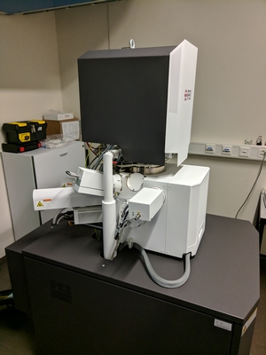
The FEI Apreo Volumescope is a scanning electron microscope equipped with a built in microtome, which is used for 3D serial block face imaging.
The Apreo is operated by the MAPS Software, which allows the automated acquisition of high resolution images from large areas in X Y and Z direction (10x10x 10 nm).
The system allows the imaging of larger areas than our focused ion beam scanning electron microscope.
MAPS is a modular software application for automated acquisition of high resolution images from large areas. (Navigate, Tile, Stitch, Correlate and Analyse Data)
MAPS allows you to:
- Acquire high resolution images over large areas
- Easily find regions of interest
- Acquire images from a sample with different settings
- Correlate data from different sources
There is a free Offline MAPS Viewer on fei.com/software/maps/ and on our virtual machines (VM).
Location
University Zurich, Irchel Campus, Room Y42-F-54.
Training Request
Follow this link to apply for an introduction to the microscope.
Technical Specifications
Electron column
field emssion electron source (Schottky-Emitter)
Acceleration voltage: 500 V to 30kV.
Detector Systems
Inlens: T1, T2, T3
Onlens: detachable low vacuum detector (VS-DBS)
Chamber: Everhart-Thornley type SE detector
Chamber: LVD low vacuum SE detector
Chamber: STEM detector for TEM like imaging
Chamber: retractable backscattered electron detector
In-chamber microtome (volumescope)
The microtome is able to cut sections between 40 nm and 100 nm thickness in the chamber.
The acquired volume can be up to 500 μm x 500 μm x 500 μm large.
Accessories
Stage: 5-axis, fully motorized stage.
Sample holder for TEM grids for STEM - mode
System controls
FEI control software.
MAPS Software
Literature and Links
Responsible Persons
If you have questions about the device please contact the responsible person.
Make sure to acknowledge the Center for Microscopy in your publication to support us.
How to acknowledge contributions of the Center for Microscopy