CLSM - Aberrior INFINITY LINE STED upright (Irchel)
Abberior INFINITY STED
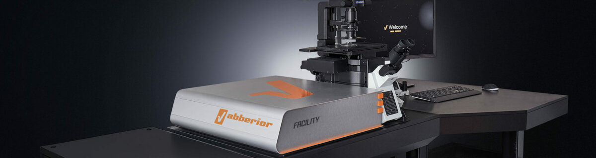
The Abberior INFINITY STED (STimulated Emission Depletion) microscope, a state-of-the-art super-resolution and confocal microscopy tool, is designed around an upright BX63F microscope body. It is equipped with four LED lines for widefield fluorescence, four laser lines, and one additional depletion laser lines. This advanced setup utilizes the STED principle to facilitate imaging that surpasses the diffraction limit. Under optimal conditions, the instrument can achieve a resolution of 42 nm laterally (xy) and 75 nm axially (z). Enhancements include a special MATRIX array detector, which improves the signal-to-background ratio, resolution, and dynamic range. With its adaptive optics for aberration correction, the microscope preserves resolution and brightness deep within thicker samples. Furthermore, Adaptive Illumination technology allows for the highest resolution and live-cell super-resolution imaging at ultra-low light levels, offering versatile and powerful imaging capabilities.
Lateral resolution (XY): 42 nm, axial resolution (Z): 75nm (typical experimental FWHM values with UPLAXPO60XO Oil immersion objective lens, NA 1.42, subresolution Crimson beads of 40 nm diameter and excitation at 640nm, 775 nm STED 100% of 3W)
Responsible Person |
|
Location |
University Zurich, Irchel Campus, Room Y11J24 |
Request a training |
Follow thislink to apply for an introduction to the microscope. |
Technical Specification
Microscope |
|
||||||||||||||||||||||||||||||||||||
Light Sources and Lasers |
|
||||||||||||||||||||||||||||||||||||
Scanner |
|
||||||||||||||||||||||||||||||||||||
Available Optics |
|
Additional Features
RAYSHAPE |
RAYPSHAPE (adaptive optics) system uses a deformable mirror to correct aberrations in microscopy imaging, particularly in inhomogeneous specimens like tissue. This technology dynamically adjusts the mirror's surface to realign distorted light rays, ensuring sharp and high-resolution images throughout different depths of the sample. It outperforms traditional correction methods by maintaining focus and image quality autonomously and continuously as the microscope examines various depths. |
MATRIX detector |
The MATRIX detector by Abberior Instruments enhances microscopy by using numerous photodiodes on a single chip, viewing samples from various angles to improve resolution and reduce background noise. It features multiple avalanche photo diodes (APDs) with high quantum efficiency, arranged hexagonally. This configuration significantly reduces background levels in STED images, enhancing clarity and detail. The multiple detection points enable precise foreground-background distinction without relying on artefact-prone mathematical algorithms, improving optical sectioning and data analysis. |
TIMEBOW |
TIMEBOW is a sophisticated tool designed for fluorescence lifetime imaging, applicable in both confocal and STED microscopy. It excels in differentiating fluorescence lifetimes due to varying types of autofluorescent molecules and their nano-environments. The system optimizes image quality by integrating MATRIX detection to remove background, allowing for selective exclusion of STED-affected emission and improvement in resolution. TIMEBOW also enables the distinction of dyes with similar excitation properties by using lifetime information, which is particularly useful for complex samples where multiple fluorophores are present. This capability allows for detailed, multi-color imaging with enhanced resolution, using a single STED laser. |
FLEXPOSURE |
Adaptive illumination dramatically reduces the light dose on the sample by up to two orders of magnitude through decreasing or shutting down laser irradiation, whenever it is clear that there is no structure present at the current pixel. This substantially reduces photobleaching. Often, FLEXPOSURE makes all the difference between a crisp image and no superresolution image at all. |
Labeling for STED microscopy
| Labelling Protocols | A good first step is to start with a protocol that's already known to work well for confocal microscopy. It's likely that it will be just as effective for STED microscopy, especially if you swap out the dyes for ones that are compatible with STED. | |||||||||
|---|---|---|---|---|---|---|---|---|---|---|
| Coverslips | It's advised to use glass coverslips that are precisely 170 um thick (#1.5 or #1.5H). This thickness is optimal as most objective lenses on Abberior systems are calibrated for it. Please avoid using plastic coverslips or chambers with a plastic coverslip base, as plastic can lead to aberrations and polarization alterations in the STED beams, adversely affecting the quality of the imaging. | |||||||||
| Embedding Media |
|
|||||||||
| Dyes for STED Microscopy |
*For STED imaging, please use organic dyes whenever possible. Organic dyes are superior to fluorescent proteins (FPs), especially with respect to brightness and photostability. As an alternative to FPs in live-cell experiments, cell-permeable organic dyes (e.g. SiR or ATTO590) may be used in conjunction with self-labelling protein tags e.g. SNAP- and/or Halo-tag.
|
|||||||||
| Multi colour recommendation |
|
Make sure to acknowledge the Center for Microscopy in your publication to support us.
How to acknowledge contributions of the Center for Microscopy