CLSM - Leica SP8 inverse (Gloriastrasse)
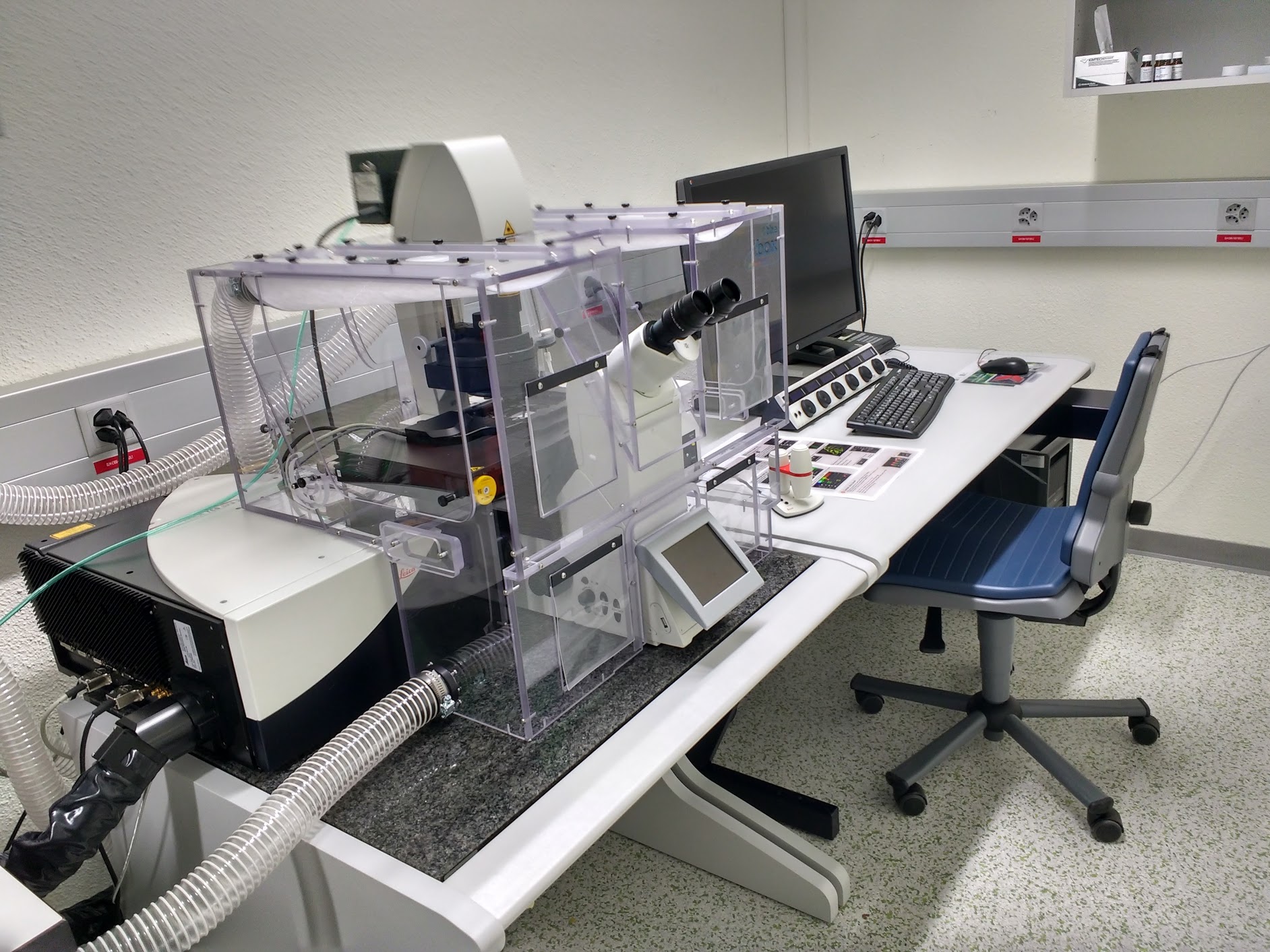
The Leica SP8 is an automated inverted confocal laser scanning microscope allowing simultaneous acquisition of 5 fluorescent channels and 1 transmitted light channel (BF or DIC). Further, the system is outfitted with a very flexible white light laser source. A climate chamber and resonant scanner favor confocal live cell studies.
Location
University Zurich, Gloriastrasse 30, Room GLM F01.
Training Request
Follow this link to apply for an introduction to the microscope.
Technical Specifications
Microscope
- inverted confocal microscope (Leica DMi8 CS with AFC, Model SP8)
- 5 freely selectable emission detection windows
- motorized x/y stage
- 3D capability
- time lapse capability
Light Sources and Lasers
- Halogen lamp for transmitted light
- External fluorescence lamp LQ-HXP 120
- Diode Lasers: 405 nm (50mW), 442nm
- Argon Laser (65mW): 458 nm, 476 nm, 488 nm, 496 nm, 514 nm
- White light laser (WLL): continuous laser output in the range of 470-670nm; AOTF allows to choose up to 8 different wavelengths simultaneously; pulsed laser system with a repetition rate of 80 MHz
Scanners (tandem scanner system)
- confocal point-scanning - optical sectioning
- zoom function
- scan field rotation
- Regular scanner (field-of-view scanner): 1 Hz to 1800 Hz
- Resonant scanner: 8 kHz
Detectors
- 5 confocal fluorescence channels: one Photomultipliertubes (PMTs) and four super-sensitive Hybrid detectors (HyD)
- one transmission light-channel (PMT-Trans)
Camera System
- Hamamatsu Orca Flash 4.0 V3 high sensitivity, digital monochrome scientific cooled sCMOS camera
- QE > 82% @ 560 nm
- 2048 x 2048 pixel (6.5 um x 6.5 um)
- 16 bit dynamics
Objectives
| Name | Magnification | NA | Immersion | WD (mm) |
|---|---|---|---|---|
| HC PL FLUOTAR | 10x | 0.3 | Air | 11.0 |
| HC PL APO CS2 | 20x | 0.75 | IMM | 0.68 |
| HC PL APO | 40x | 0.95 | Air | 0.17 |
| HC PL APO CS2 | 63x | 1.4 | oil | 0.1 |
| HC PL APO CS2motCorr | 63x | 1.2 | water | 0.3 |
CS2 objectives: improved color correction, perfect VIS-405; IMM = multi immersion (either water, glycerol or oil)
Fluorescence Filters
Filter for Epi-Fluorescence using the Filter turret
| Name | Excitation Range | Excitation Filter | Dichroic | Emission Filter |
|---|---|---|---|---|
| DAPI | UV | BP 350/50 | 400 | BP 460/50 |
| GFP | blue | BP 470/40 | 495 | BP 525/50 |
| RHOD | green | BP 546/10 | 560 | BP 585/40 |
| QUAD-S | UV/blue/green/red | LP (see fast filter wheels) | 425; 505; 575; 660 | LP (see fast filter wheels) |
Filter for Epi-Fluorescence using the QUAD-S in combination with the fast filter wheels
| Filter Name | Excitation/Emission Range | Filters/Bands |
|---|---|---|
| DFTC-EX | UV/blue/green/red | BP 350/50, BP 490/20, BP 555/25, BP 645/30, EMPTY |
| DFTC-EM | UV/blue/green/red | BP 455/50, BP 525/36, BP 605/52, BP 705/72, EMPTY |
Accessories
- controlled environmental conditions (CO2, temperature, humidity)
- Matrix screening software (reservation needed)
Literature and Links
ibiology - Optical Sectioning and Confocal Microscopy
Leica Science Lab
Responsible Persons
If you have questions about the device please contact the responsible person.
Make sure to acknowledge the Center for Microscopy in your publication to support us.
How to acknowledge contributions of the Center for Microscopy