CLSM - Nikon A1R+ (Balgrist Campus)
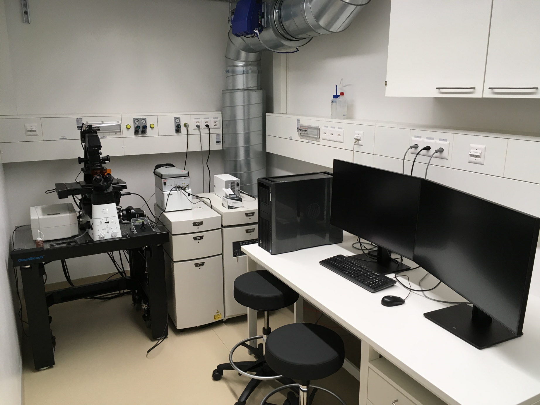
The Nikon A1R+ is an automated inverse confocal laser scanning microscope allowing simultaneous acquisition of 5 fluorescent channels and 1 transmitted light channel (BF). In addition, the system can be operated as a widefield fluorescent microscope. The system is ideal for fixed samples.
Location
University Zurich, Balgrist Campus, Lengghalde 5, 8008 Zurich, Room Microscopy W.112.
Training Request
Follow this link to apply for an introduction to the microscope.
Technical Specifications
Microscope
Nikon Eclipse Ti2 inverted
Light sources and lasers
- Lumencor sola light engine
- Diode lasers: 405 nm (LD, 20 mW), 445 nm (LD; 20 mW), 488 nm (DPSS, 20 mW), 514 nm (DPSS, 20 mW), 561 nm (DPSS, 20 mW) and 640 nm (LD, 20 mW)
LD = Laser diode, DPSS = diode pumped solid state
Camera System
cooled Nikon monochrome fluorescent DS-Qi2 CMOS camera
4908 x 3264 pixel (16.25 MP)
pixel size 7.3 um
25 mm FOV
Frame rate: 6 fps (4908×3264) up to 45 fps (1636×1088)
Scanners
Dual scanning system:
Regular Galvano scanner:
- allows to capture up to 5 channels simultaneously (4x florescent and 1x transmitted light)
- Speed: Scanner: galvano scanner x2
- Pixel size: max. 4096 x 4096 pixels
- Scanning speed:
- Standard mode: 2 fps (512 x 512 pixels, bi‐direction), 24 fps (512 x 32 pixels, bidirection)
- Fast mode: 10 fps (512 x 512 pixels, bi‐direction), 130 fps (512 x 32 pixels, bidirection)
- Zoom: 1‐1000x continuously variable with “best fit” button as a time saving method of acquiring the maximum zoom before empty magnification (Nyquist criteria).
Resonant Scanner:
- allows to capture up to 5 channels simultaneously (4x florescent and 1x transmitted light)
- Speed: Scanner: resonant scanner (X‐axis, resonance frequency 7.8 kHz), galvano scanner (Yaxis)
- Pixel size: max. 1024 x 1024 pixels
- Scanning speed: 30 fps (512 x 512 pixels) to 420 fps (512 x 32 pixels), 15,600 lines/sec (line speed)
- Zoom: 7 steps (1x, 1.5x, 2x, 3x, 4x, 6x, 8x)
- Acquisition method: Standard image acquisition, High‐speed image acquisition, Simultaneous photoactivation and image acquisition
Objectives
| Name | Magnification | NA | Immersion | WD (mm) |
|---|---|---|---|---|
| Plan Apo l | 2x | 0.1 | Air | 8.5 |
| Plan Fluor DIC L N1 | 10x | 0.3 | Air | 16.0 |
| Plan Apo l | 20x | 0.75 | Air | 1.0 |
| Plan Fluor DIC M N2 | 40x | 0.75 | Air | 0.66 |
| Plan Apo Ph2 DM | 60x | 0.95 | Air | 0.2 |
| Plan Apo l | 60x | 1.4 | Oil | 0.13 |
Ph = Phase contrast, DIC = differential interference contrast
Detector System
Nikon GaAsP 4PMT filter-based Detector unit equipped with two GaAsP PMTs (channel 2 & 3) and two multialkali PMTs (channel 1 & 4).
Filter cubes are organized in three filter turrets (6 positions in each). The detector unit comes with the following filter sets:
- 450/50 (1st filter wheel)
- 482/35 (2nd filter wheel)
- 525/50 (2nd filter wheel)
- dual BP 595/50 and 700/75 (3rd filter wheel)
- 540/30 (3rd filter wheel).
Fluorescence Filters for widefield
| Name | Excitation Range | Excitation Filter | Dichroic | Emission Filter |
|---|---|---|---|---|
| DAPI | UV | BP 377/50 | 409 | BP 447/60 |
| GFP | blue | BP 446/40 | 495 | BP 525/50 |
| YFP | green | BP 500/24 | 520 | BP 542/27 |
| CY3 | orange | BP 531/40 | 562 | BP 593/40 |
| CY5 (not installed - optional) | red | BP 628/40 | 660 | BP 692/10 |
Available Accessories
- IR Hardware Autofocus (Nikon Perfect Focus System - PFS)
Responsible Persons
If you have questions about the device please contact the responsible person.
Make sure to acknowledge the Center for Microscopy and Image Analysis in your publication to support us.
How to acknowledge contributions of the Center for Microscopy and Image Analysis