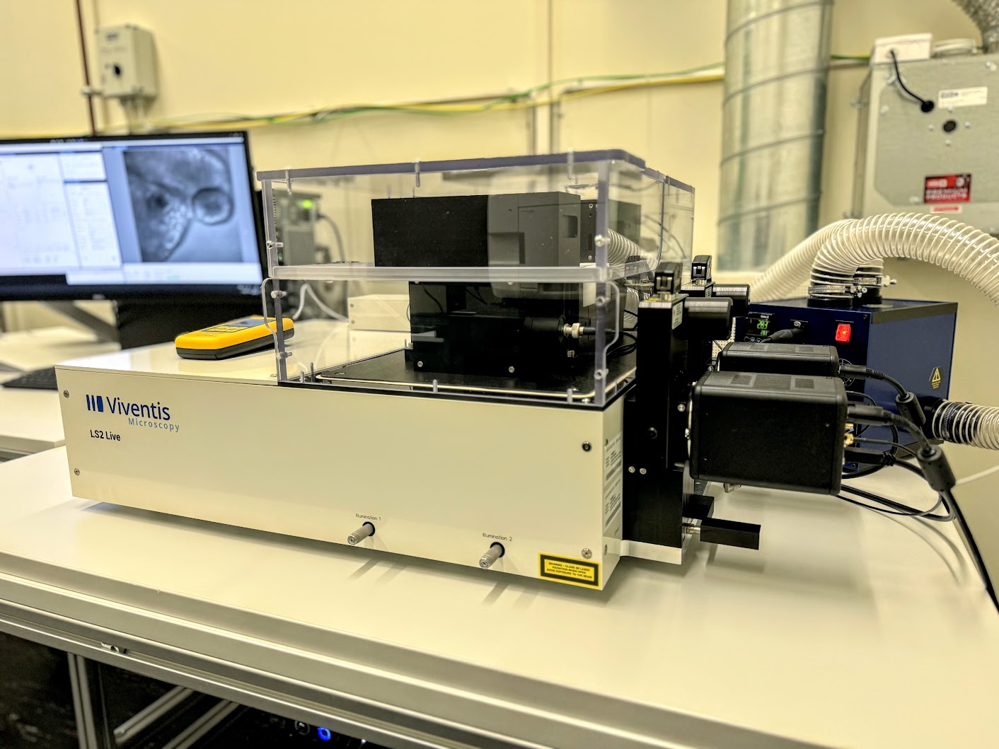
Lightsheet - Leica Viventis LS2 Live
The Lieca Viventis LS2 Live is an open-top light sheet microscope, equipped with dual illumination and dual detection capabilities, tailored for delivering volumetric imaging of large samples. This state-of-the-art system will soon be available at the Center for Microscopy and Image Analysis. Its versatility makes it an optimal choice for various live imaging applications, including the observation of organoids and embryos. Furthermore, this cutting-edge technology streamlines multiplex light-sheet microscopy, offering enhanced functionality.
Location
University Zurich, Irchel Campus, Room Y42-H-91.
Training Request
Follow this link to apply for an introduction to the microscope.
Technical Specifications
Excitation path
Light Sheet
Light sheet generated by scanning of a Gaussian beam.
- Double illumination using two Nikon 10x obkectives 0.2 NA
- Three switchable light-sheets with thicknesses (FWHM) of approximately 2, 3 and 7 μm for different sample sizes.
- Automatic position-specific light-sheet alignment.
Lasers
External laser combiner, Omicron LightHUB+.
- Module 488nm (60mW)
- Module 561nm (60mW)
- Module 638nm (60mW)
Transmitted Light Illumination
LED source
Detection path
Single magnification 25x (FOV 590 µm), pixel size-XY 260 nm
Objectives
- Two Nikon 25XW NA 1.1 water immersion ojectives.
Cameras
- Two high sensitivity Hamamatsu ORCA-Fusion cameras.
Emission Filters
- Semrock 525/50-25
- Semrock 488/568-25
- Quadband Chroma 405/488/561/640
Sample Holder
Dedicated multi-well sample holder
- Samples are held in sample chamber of custom shapes adaptable to the sample being imaged.
- Easy sample mounting from top and medium exchange possible during running time-lapse .
- Sample can be placed in different media (e.g drugs vs control experiments).
- You can ask for other holder options that can be adjusted to your samples' needs.
Recent Publications
2023
- Harasimov et al., Actin-driven chromosome clustering facilitates fast and complete chromosome capture in mammalian oocytes Nature Cell Biology
- Olivetta et al., The nuclear to cytoplasmic ratio drives cellularization in the close animal relative Sphaeroforma arctica bioRvix 2022 • Ozelci et al., Deconstructing body axis morphogenesis in zebrafish embryos using robot-assisted tissue micromanipulation. Nature Communications
2022
-
Ozelci et al., Deconstructing body axis morphogenesis in zebrafish embryos using robot-assisted tissue micromanipulation. Nature Communications
-
Ishihara et al., Topological morphogenesis of neuroepithelial organoids. Nature Physics
-
de Medeiros et al., Multiscale light-sheet organoid imaging framework Nature Communication
-
Naganathan et al., Left-right symmetry of zebrafish embryos requires somite surface tension. Nature
-
So et al., Mechanism of spindle pole organization and instability in human oocytes. Science
Further information (internal UZH use only)
Follow this link for further background information, documents and links.
Responsible Persons
If you have questions about the device please contact the responsible person. For training requests register here.
Make sure to acknowledge the Center for Microscopy in your publication to support us.
How to acknowledge contributions of the Center for Microscopy
