Navigation auf uzh.ch
Navigation auf uzh.ch
This inverted wildfield system is suited to image live cells as well as fixed samples in fluorescence and transmission light modes. A temperature controlled environment is possible for live cell experiments. The system does not have CO2 control. It is well suited for phase contrast imaging of bacteria and does allow complex imaging tasks.
University Zurich, Irchel Campus, Room Y38-M-77.
Follow this link to apply for an introduction to the microscope.
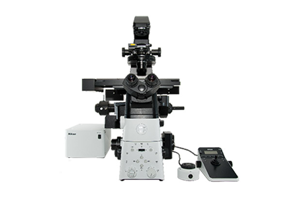
Microscope
Nikon Eclipse Ti2-E fully motorized microscope:
- Motorized encoded xy-stage and motorized encoded z-drive
- Perfect Focus System 4
- Motorized fluorescence turret, filter-wheel, condenser turret, light path switcher
- Lumencor SpectraX with 6 individual LED-lines (395, 440, 470, 510, 550/575, and 640)
| Name | Magnification | NA | Immersion | WD (mm) |
|---|---|---|---|---|
| CFI Plan Apochromat Lambda | 2x | 0.1 | Air | 8.5 |
| CFI Plan Apochromat Lambda DIC | 10x | 0.45 | Air | 4.0 |
| CFI Plan Apochromat Lambda PH2 | 20x | 0.75 | Air | 1.0 |
| CFI Plan Apochromat Lambda DIC | 40x | 0.95 | Air | 0.25 |
| CFI Plan Apochromat Lambda PH2 | 40x | 0.6 | Air | 3.6 |
| CFI Plan Apochromat Lambda PH3 | 100x | 1.45 | Oil | 0.13 |
Ph = phase contrast, DL = Dark Low Contrast, DM = Dark Medium Contrast, DIC = Differential Interference Contrast
| triple filter cube CFP/YFP/mCherry | Excitation | Mirror | Emission |
|---|---|---|---|
| 426-450 | 464-486 | 464-486 | |
| 497-519 | 532-554 | 532-554 | |
| 567-588 | 603-800 | 603-800 | |
| quadruple filter cube Dapi/FITC/TRITC/Cy5 | |||
| 352-404 | 414-450 | 414-450 | |
| 461-487 | 499-530 | 499-530 | |
| 543-566 | 580-611 | 580-611 | |
| 626-644 | 659-800 | 661-800 |
wavelength in nanometer
| Emission filter wheel | Emission |
|---|---|
| 414-450 | |
| 499-530 | |
| 580-611 | |
| 662-732 |
wavelength in nanometer
Orca Fusion CoaXpress sCMOS
2304 x 2304 pixel (6.5 x 6.5um pixel size)
Responsible Persons
If you have questions about the device please contact the responsible person.
Make sure to acknowledge the Center for Microscopy and Image Analysis in your publication to support us.
How to acknowledge contributions of the Center for Microscopy and Image Analysis