Header

Search

Leica LMD7

Leica LMD7 Laser microdissection

Make sure to acknowledge the Center for Microscopy and Image Analysis in your publication to support us.

How to acknowledge contributions of the Center for Microscopy and Image Analysis

The Leica LMD7 is a laser microdissection system for isolating selected tissue regions, cell populations or individual cells for downstream molecular analysis. It combines brightfield and fluorescence microscopy with a precisely controlled UV laser, allowing samples to be selected according to their morphology or fluorescent labelling. In the standard collection workflow, dissected material falls by gravity into a collection vessel below the sample, enabling contact-free collection for applications in genomics, transcriptomics and proteomics.

lmd
Zoom
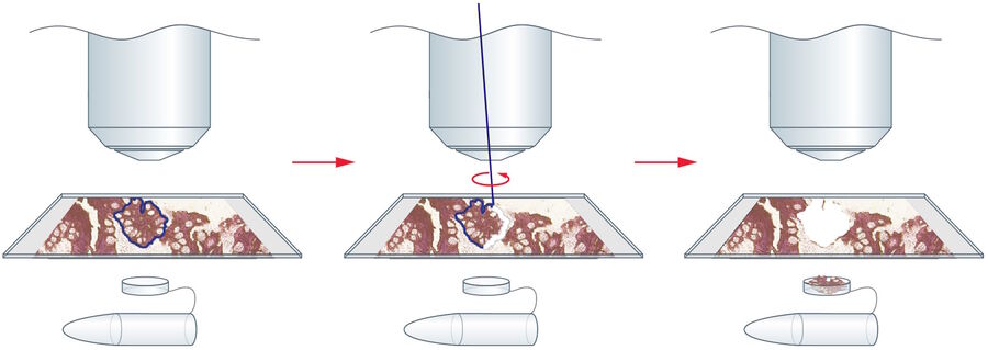
The principle of laser microdissection based on the Leica LMD system: Step 1) Defining the region of interest; Step 2) Steering the laser beam with the optics along the cut line; and Step 3) Collecting the specimen with gravity.

Responsible Person

responsible person

Location

University Zurich, Irchel Campus, Room Y42-H-83. 

Training Request

Follow this link to apply for an introduction to the microscope

Applications

  • Isolation of defined tissue regions, cell populations and individual cells from heterogeneous samples.
  • Selection of targets using histological staining or fluorescence labelling.
  • Sample preparation for DNA and RNA analysis, including PCR and sequencing.
  • Sample preparation for proteomics, including spatially targeted mass spectrometry.
  • Dissection of compatible cell cultures for downstream analysis or subsequent cultivation, depending on the sample and preparation method.
Modalities

Laser Microdissection


A focused UV laser cuts around user-defined regions of interest. The laser beam is steered through the optics while the specimen remains stationary during cutting within the field of view. Laser settings can be adjusted to suit different sample types and thicknesses. Selected regions can be outlined before cutting or dissected interactively in real time.

Brightfield and Fluorescence Imaging


Transmitted-light brightfield imaging allows the identification of tissue morphology and histological staining. Widefield fluorescence imaging supports the identification and dissection of labelled structures using DAPI, GFP, Alexa Fluor 594 and Cy5 filter sets.

Cell Culture Dissection

Compatible membrane-based culture vessels allow selected cells or cell clusters to be isolated for molecular analysis or subsequent cultivation. Sample substrates, collection media and environmental requirements should be discussed with the responsible person before planning an experiment.


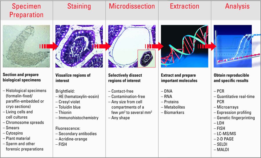
The overall process required for laser microdissection, from specimen preparation to analysis, is shown above (source: Leica)

Technical Specifications

Microscope Body

  • Upright Leica DM6 B research microscope with motorised Z focus.
  • Motorised seven-position objective turret.
  • Motorised five-position fluorescence filter turret.
  • Internal Filter Wheel, Excitation Manager and Fluorescence Intensity Manager.
  • DIC-capable LMD condenser with S40/0.40 condenser head.
  • Collection-cap inspection hardware for checking collected material.

Light Sources and Laser

  • LED illumination for transmitted-light microscopy.
  • CoolLED pE-300 white MB fluorescence illumination with spectral coverage from DAPI to Cy5 excitation.
Laser specification Configuration
Laser type Diode-pumped solid-state UV laser
Wavelength 349 nm
Maximum pulse energy 120 µJ
Adjustable pulse frequency 10-5,000 Hz
Adjustable parameters Laser power, pulse frequency, aperture and offset
Beam positioning Optical steering of the laser beam


Camera System

  • Leica K7 colour CMOS camera with 7.1 megapixels.
  • Sony IMX420 sensor, 3,200 × 2,200 pixels.
  • Sensor pixel size: 4.5 × 4.5 µm.
  • 8-, 12- and 16-bit acquisition modes.
  • Exposure range: 1 ms to 30 s.
  • Up to 110 frames per second at full resolution in streaming mode, depending on bit depth, exposure time and workstation performance.
  • Hardware triggering and 10 Gigabit Ethernet interface.

Motorised Stage


The LMT350 Ultra scanning stage provides independent positioning of the specimen and collection vessel. The standard specimen holder accommodates four microscope slides.

Specification Specimen stage Collector stage
Travel range 120 × 80 mm 100 × 112 mm
Repeatability Within ±0.5 µm Within ±5 µm
Positioning accuracy Within ±2 µm Within ±10 µm
  • Linear motor drives with a maximum travel speed of 200 mm/s.
  • Absolute position measurement with an encoder resolution of ≤5 nm.
  • Multiwheel controller for X, Y and Z adjustment.

Collection Formats


Collection format Configuration
Individual PCR tubes Up to four 0.2 or 0.5 ml tubes with flat caps
Strip caps 8- and 12-position tube-strip caps
PCR plates Non-skirted or semi-skirted 96-well PCR plates
Cell culture collection 96-well collector for compatible live-cell culture workflows
Direct-contact pickup Special 1.5 ml tubes with filled caps

Gravity collection is contact-free. The dedicated 1.5 ml tube option uses direct contact to pick up material.

Available Optics


Objective Magnification NA Immersion Working distance (mm)
Plan 4× 0.10 Air 26.2
UVI Microdissection 5× 0.12 Air 13.5
HC PL FLUOTAR 10× 0.30 Air 11.0
HC PL FLUOTAR L CORR 20× 0.40 Air 6.9
HC PL FLUOTAR L CORR XT 40× 0.60 Air 3.3-1.9
HC PL FLUOTAR L CORR XT 63× 0.70 Air 2.6-1.8

The 20×, 40× and 63× objectives have correction collars. The 5× UVI objective and the 40× and 63× XT objectives are specifically designed or optimised for laser microdissection.

Available Filters


Filter set Fluorescence channel
LMD-DAPI ET Blue
LMD-GFP ET Green
LMD-Alexa594 Red
LMD-Cy5 ET Far-red

The GFP filter set has a 470/40 nm excitation filter, a 495 nm dichroic and a 525/50 nm emission filter. The LMD fluorescence filter sets allow UV laser cutting during fluorescence observation.


Environmental Protection

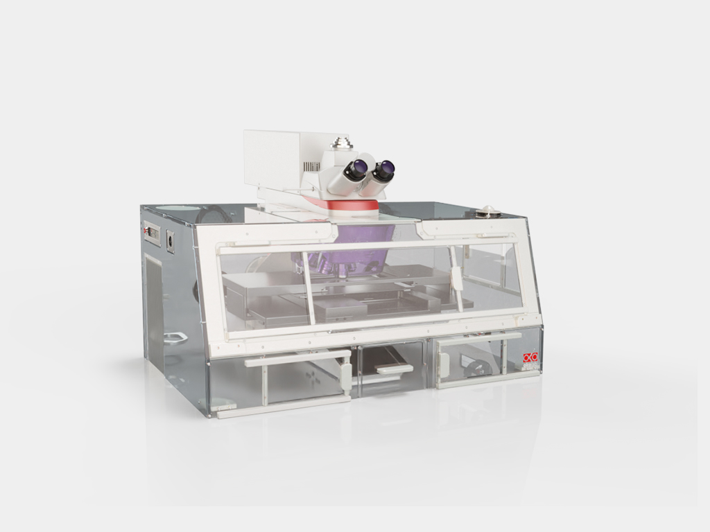
An Okolab H201-Enclosure-LMD transparent enclosure shields the specimen area from airflow and environmental contamination. This configuration provides a protective enclosure without active temperature or CO₂ control.

Accessories

  • Holder for four standard microscope slides.
  • Inlays for large slides, Petri dishes, 18-well ibidi slides and slide stacks.
  • Frame support for Leica LMD FrameSlides.
  • Collection holders for PCR tubes, strip caps and 96-well plates.
  • Passive antivibration table.
LMD
Zoom


Software

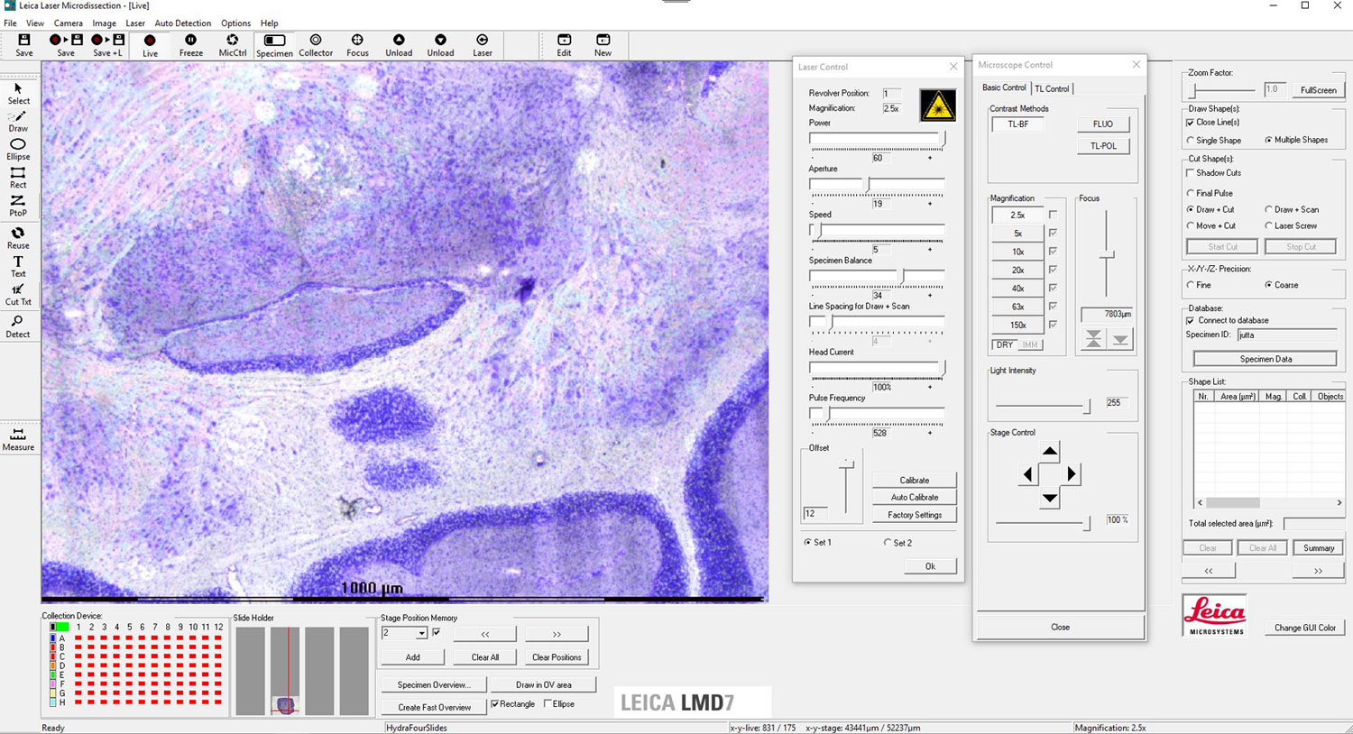
  • Leica LMD Application Software for microscope and laser control, region selection, cutting and collection.
  • Draw & Cut and Move & Cut workflows, saving and loading cutting shapes, and sample overview imaging.
  • LMD Database and LIF-Browser for documentation and review of microdissection experiments.
  • HP workstation with Windows 11 Pro, 32 GB RAM, a 4 TB data SSD and a 31.5-inch UHD monitor.
lmd
Zoom


Sample Requirements

  • Suitable samples include FFPE sections, frozen sections and compatible cell culture preparations.
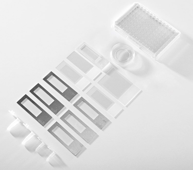
  • For standard gravity collection, prepare tissue sections on suitable laser-microdissection membrane slides. Slide and membrane selection depend on the sample and downstream analysis.
  • Agree on section thickness, fixation and staining before sample preparation. These affect target identification, cutting, collection and the quality of extracted DNA, RNA or proteins.
  • Keep the area intended for dissection accessible. Discuss coverslipping and mounting media with the responsible person before mounting samples.
  • For cell culture experiments, use compatible membrane-based vessels and discuss media, collection and environmental requirements in advance.

User Guide

TBA

Links & Literature